Monday, December 10, 2007

Biochemical tests for bacteria indentification-Group post 2

Biochemical tests for gram negative bacteria identification

1)Indole

-measure the ability to hydrolyse and deaminate tryptophan

-Klesiella-enterobacter-salmonella-serratia are mostly negative

-positive-red colour


2)Methyl red

-methyl red, a pH indicator with a range between 4.4(red) and 6.0(yellow)

-only species that produce suffiicient acids can maintian the pH at below4.4 against the buffer system of the test medium

-most species of Enterobacteriaceae produce strong acids. Enterobacter-serratia do not produce enough acids

-positive-stable red colour in the surface layer of the medium


3)Voges-proskauer reaction test

-this test is based on the conversion of acetoin to a red coloured complex through the action of KOH, atmospheric 02 and alpha napthol

-Klesiella-enterobacter-serratia is able to perform this pathway

-red colour at the surface of the medium after 15 mins following the addition of reagents


4)Citrate utilisation test

-some bacteria have the ability to utilize citrate as the sole carbon sourc and turn the medium allkaline due to production of ammonia

-Escherichia-Edwardisella-shigella-salmonella cannot utilise citrate as the sole source of carbon

-positive-from colour green to blue


5)Urease test

-some species posses the enzyme urease and able to hydrolyze urea with the release of ammonia and carbon dioxide

-this is used mainly to differentiate urease positive Proteus species from other member of Enterobacteriaceae

-positive-yellowish orange to pink


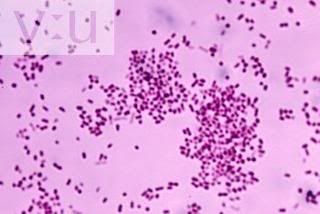
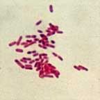
Biochemical tests for gram positive bacteria


1)Oxidase test

-this is to differentiate those that possess the enzyme cytochrome oxidase c from those that lack of the enzyme

-useful in screening for bacteria species which belong to the Enterobacteriaceae or the Pseudomonas genus

-positive-development of purple colour


2)Coagulase test

-the coagulase test is used to differentiate staphylcoccus aures from other staphylcoccus species

-positive-clot forms

Particulars of patient

Name:Khong Fay Seah
Sex:Female
Age:27
Clinical Diagnosis
Complaints: fever,chills,dysuria
Diagnosis:urinary tract infection
Antibiotic treatment: if any (nil)


Possible Microorganisms

Description

Investigations

Escherichia coli(Most common cause of UTI)


-Belongs to family of Enterobacteriaceae(Escherichia)

-Clinical findings: UTI (Pyelonephritis and Cystitis), diarrhea disease

-Is a member of normal intestinal flora

-Microscopy and gram staining: Gram negative rod

-Culture: Haemolyic colonies on blood agar, lactose fermenting colonies (Red/Pink) on MacConkey agar, green metallic sheen on EMB agar

-Biochemical tests:
-Indole positive
-Methyl red positive
- Voges-Proskauer (VP) negative
-Urease negative
-Citrate negative
-Catalase positive
-Oxidase negative
-TSI: acidic slant/acidic deep/no H2S

-Susceptibility testing: ampicillin, cephalosporin, aminoglycosides, sulfonamides

Staphylococcus saprophyticus(Second most common cause of UTI)


-Belongs to family of staphylococci

-Clinical findings: Urinary tract infection

-Present in the urinary tract and bladder of sexually active females

-Microscopy and gram staining: Gram positive cocci in clusters

-Culture: Pale colonies on blood agar

-Biochemical tests:
-Catalase positive
-Coagulase negative

-Susceptibility testing:Quinilone

Klebsielle Pneumoniae


- Belongs to family of Enterobacteriaceae(Klesbsiella)

- Clinical findings: urinary tract infection

-Present in the respiratory tract and feces of about 5% normal individuals

-Microscopy and gram staining: Gram negative rod

-Culture: Pink, mucoid colonies on MacConkey agar

-Biochemical tests:
-Indole negative
- Methyl red negative
-Voges-Proskauer reaction positive
- Urease negative
-Citrate positive
- TSI: acidic slant/acidic deep/no H2S

-Susceptibility testing:Cephalosporin

Enterobacter aerogenes


- Belongs to family of Enterobacteriaceae(Enterobacter)

- Clinical findings: urinary tract infections and sepsis

- Present in the intestinal tract-

-Microscopy and gram staining: Gram negative rod

-Culture:Pink to purple colonies on EMB agar

-Biochemical tests:
-Indole negative
-methyl red negative
-VP positive
-urease negative
- Citrate positive
-TSI: acidic slant/acidic deep/no H2S

Susceptibility testing: cephalosporin

Proteus mirabilis


- Belongs to family of Enterobacteriaceae(Proteus)

- Clinical findings: urinary tract infections and produce bacteremia, pneumonia

-Microscopy and gram staining: Gram negative rod
-Culture: Pale colonies on MacConkey agar, swarming growth on blood agar

-Biochemical testing:
-Indole negative
-Methyl red positive
-VP positive/negative
-Urease positive
- Citrate positive
-Oxidase negative
-TSI: alkaline slant/acidic deep/H2S

-Susceptibility testing: penicillin, cephalosporins, quinolones

Enterococcus faecalis


- Belongs to family of Streptococci

-Clinical findings: abdominal abscess, urinary tract infection ((bladder infection, Pyelonephritis)

-Present in colon

-Microscopy and gram staining: Gram positive cocci in chains

-Culture: Red dot colonies on MacConkey agar

-Biochemical testing:
-Decolourise the litmus milk,
-Indole negative
-Catalase negative

-Susceptibility testing: Vancomycin,ampicillin

Pseudonomas aeruginosa


- Belongs to family of Pseudomonads

-Clinical findings: Typically infects the pulmonary tract, urinary tract(Pyelonephritis)

-Distributed in nature and is commonly present in moist environments in hospital

-Microscopy and gram staining: Gram positive spherical

-Culture: Greenish pyocyanin pigment on MacConkey agar

-Biochemical testing:
-Indole negative
-Urease negative
-Oxidase positive,
-TSI: allaline slant/alkaline deep

-Susceptibility testing: Ciprofloxacin,imipenam


PICTURES OF THE POSSIBLE CAUSATIVE AGENTS


Photo Sharing and Video Hosting at Photobucket


E Coli ( red dot colonies on Mac agar)

Photo Sharing and Video Hosting at Photobucket

K. Pneumonia (red/pink colonies on Mac)

Photo Sharing and Video Hosting at Photobucket

Staphyloccocus Saprophyticus
Photo Sharing and Video Hosting at Photobucket
Pseudomonas aeruginosa
Photo Sharing and Video Hosting at Photobucket
Enterococcus faecalis
Photo Sharing and Video Hosting at Photobucket
Enterobacter aerogenes
Photo Sharing and Video Hosting at Photobucket
Proteus mirabillis
References

http:www.yahoo.com>image search> Proteus mirabilis
http:www.yahoo.com>image search> Enterobacter Aerogenes
http:www.yahoo.com>image search>Enterococcus faecalis
http:www.yahoo.com>image search>Pseudomonas aeruginosa
http:www.yahoo.com>image search>Staphyloccocus Saprophyticus
http:www.yahoo.com>image search>K. Pneumonia
http:www.yahoo.com>image search> E Coli
http://www.mc.maricopa.edu/~johnson/labtools/Dbiochem/emb.html>EMB agar
http://en.wikipedia.org/wiki/MacConkey_agar> MacConkey Agar
http://en.wikipedia.org/wiki/Blood_agar#Blood_agar_types> blood agar
Book
Tony Hart, Paul Shears (2004).Color Atlas of Medical Microbiology: Elsevier’s Health Sciences Department


kai lin
0503211E

Sunday, December 9, 2007

MMIC dPBL Case 3 (Maisy Hong - 2nd posting cont)

Pictures of possible causative agents of case 3

Pseudomonas aeruginosa growing on XLD agar Acinetobacter baumanii as seen under microscopy
Serratia marcescens growing on XLD agar
Candida albicans as seen under microscopy

Staphylococcus saprophyticus growing on nutrient agar Enterococcus faecalis as seen under scanning electron microscopy
Escherichia coli as seen under microscopy
Proteus mirabilis growing on XLD agar

Klebsiella pneumoniae as seen under microscopy
References:
All images are credited to
1) www.yahoo.com > image search > "microorganism name"
2) www.wikipedia.org > "microorganism name" > search
Information are credited to
3) Murray, P.R., Kobayashi, G.S., Pfaller, M.A., Rosenthal, K.S. (1994). Medical Microbiology Second Edition. London: Mosby-Year Book, Inc
4) Medical Microbiology Notes
5) www.yahoo.com > "microorganism name"
6) www.wikipedia.org > "microorganism name"
Tan Yi Wei Alex Tg02 0503222B

MMIC dPBL Case 3 (Maisy Hong - 2nd posting)

Learning issues:

1) Identify suspected microbial agents
2) Conduct self-directed learning based on specific case and symptoms
3) Propose relevant tests according to suspected causative agents
4) Propose treatment to suspected causative agents

Keywords: Female, 67 years, Fever, Chills, Bladder distension, Indwelling catheter, Urinary tract infection, Urine specimen

Patient's Particulars

Name: Maisy Hong
Age: 67 years old
Sex: Female

Clinical Diagnosis

Complaints: Fever, chills, bladder distension (inability to urinate)
Diagnosis: Urinary tract infection (UTI)
Note: On indwelling catheter (a urinary catheter is a plastic tube which is inserted through a patient's urinary tract into their bladder)

Diagnostic approach to UTI in adults

1) Patient symptomatic? Yes, go to 2a. No, go to 2b.
2a) Complicating factors? Yes, go to 3a. No, go to 3b.
2b) Asymptomatic bacteriuria
3a) Complicated UTI
3b) Recurrent episode? Yes, go to 4a. No, go to 4b.
4a) Recurrent UTI
4b) Symptoms of upper tract involvement? Yes, go to 5a. No, go to 5b.
5a) Pyelonephritis
5b) Consider cystitis, urethritis or vaginitis (lower tract)


Usually, before proceeding to microbial identification tests, a urinalysis and chemical dipstick testing will be performed on the urine sample and important factors of UTI such as leucocytes, erythrocytes, pH as well as presence of certain chemicals like nitrite are things to look out for. As we've covered this in clinical chemistry, I will just talk about the medical microbiological portion. :)


Below is a table of possible causative agents, characteristics, proposed tests for agent identification and antibiotic susceptibility testing (treatment).


(Please click the pictures for enlarged images)


Tan Yi Wei Alex Tg02 0503222B

CASE STUDY 4

Particulars of Patient
Name: Tong Wei Hong
Sex: Male
Age: 68 years old

Clinical Diagnosis
Signs and symptoms: Fever, chills, excessive phlegm, breathing problems Diagnosis: Bronchitis
Specimen collected: Sputum

Description of Bronchitis
Bronchitis is an inflammation of the bronchi (medium-size airways) in the lungs. Acute bronchitis is usually caused by viruses or bacteria and may last several days or weeks. Acute bronchitis is characterized by cough and sputum (phlegm) production and symptoms related to the obstruction of the airways by the inflamed airways and the phlegm, such as shortness of breath and wheezing. Diagnosis is by clinical examination and sometimes microbiological examination of the phlegm.

Chronic bronchitis is not necessarily caused by infection and is generally part of a syndrome called chronic obstructive pulmonary disease (COPD); it is defined clinically as a persistent cough that produces sputum (phlegm) and mucus, for at least three months in two consecutive years.

Possible Causative Agents

1. Adenovirus
Non-enveloped double-stranded linear DNA
Icosahedral nucleocapsid with a fiber protruding from each of the 12 vertices
Causes Bronchitis in the lower respiratory tract

2. Bordetella
Small, coccobacillary, encapsulated gram negative rod
Restricted to the respiratory tract (negative blood culture)

3. Haemophilus influenzae
Non-motile gram-negative coccobacillus
Found in the upper respiratory system of humans
Major cause of lower respiratory tract infections, associated with pneumonia

4. Moraxella catarrhalis
Gram-negative, aerobic, oxidase-positive diplococcus
May colonise and cause respiratory tract-associated infection in humans
Known to cause Bronchitis

5. Streptococcus pneumoniae
Gram positive lancet-shaped cocci
Arranged in pairs or short chains
Higher mortality in persons aged 65 and above


Investigations
The colour of the sputum may indicate the kind of microorganism in the sputum; yellow to green may indicate bacteria while white may indicate viruses. The sputum should be cultured to isolate the microorganisms listed above. In addition,

Biochemistry
-Gram staining
-Citrate test
-Catalase test
-Oxidase test
-Urease test

Microbiology
-Culture aerobically on blood agar plates

Antibiotic Suspectibility testing

Serology (Virus detection)
-Enzyme-Linked ImmunoSorbent Assay (ELISA)
-Haemagglutination


References
www.wikipedia.org > English > search > Bronchitis
www.wikipedia.org > English > search > Adenovirus
www.wikipedia.org > English > search > Bordetella
www.wikipedia.org > English > search > Haemophilus influenzae
www.wikipedia.org > English > search > Moraxella catarrhalis
www.wikipedia.org > English > search > Streptococcus pneumoniae


Desmond Heng
0503179D

Follow-up of case 6


Above is the procedure that i will carry out during laboratory to determine the identity of the microoraganism present in the vaginal swap.

Materials Needed:
1) Blood Agar
2) MacConkey Agar
3) Gram stain Reagent
4) Caoagulase Reagent
5) Oxidase Reagent
6) Catalase Reagent
7) Commercial ID Testing Kits
8) Biochemical Tests Reagents
9) Antibody lines
Patient: Ong Fei Fei
Age: 37 Years old
Complaints: Fever, pain during urination, virginal discharge
Diagnosis: UTI

1)Enterococcus Faecalis
Labortatory Diagnosis:
(i) Alpha, beta or non hemolytic colonies on blood agar
(ii) Grow in 6.5% NaCl and hydrolyzes esculin on the presence of 40% bile
(iii)Catalase Negative

2)Eschericahia coli
Labortatory Diagnosis:
(i) Lactose fermenting on macConkey agar
(ii) TSI show acid slant, acid butt with gas but no H2S
(iii)Indole +
(Iv) Motile

3)Enterobacter-Klebsiella-Serrtia Family
Labortatory Diagnosis:
(i) Lactose fermenting on macConkey agar
(ii) Appear as mucoid colonies on agar plate

4)Proteus-Providencia-Morganella Family
Labortatory Diagnosis:
(i) Non-lactose fermentor on Macconkey
(ii) Distinct swarming on blood agar
(iii)P.vulgaris and P.mirabilis produce H2S that blacken the butt of TSI agar
(iv) P.mirabilis is indole negative whereas the other 2 are indole positive
(v) Urease Positive

Antibiotic suceptibility Testing will be carried out on antibodies mention in previous posting after bacterial is identified.

Yeo Ching Wei
0503288C

MMIC Blog Post 2

Patient’s Particulars:
Name: Kwan Siew Yan
Sex: Female
Age: 29 years

Clinical Diagnosis:
Complaints: Diarrhea
Diagnosis: Entercolitis
Antibiotic Treatment: Nil

Possible organisms:
1) Vibrio species:
V. parahaemolyticus- Facultative anaerobes
Microscopic features:
· “Curved”, comma-shaped rods
· Motile with polar flagella
Laboratory investigations:
· Gram staining: Negative
· Oxidase: Positive on blood agar
· TSI: acid slant/acid deep without H2S production
· 0/129 (Vibriostat) Disk: Susceptible
· Nutrient Broth with 0% or 6-8% NaCl: Requires 1-7% NaCl
· Decarboxylase: Positive
· Culture media: TCBS as green colonies due to absence of sucrose fermentation
Antibiotic-susceptibility test:
· Tetracycline

2) Campylobacter species:
C. jejuni- Microaerophilic
Microscopic features:
· “S” shaped rods
· Darting motility
Laboratory investigations:
· Gram staining: Negative
· Oxidase: Positive
· Catalase: Positive
· TSI: Alkaline slant/alkaline deep
· Hippurate hydrolysis: Positive
· Culture media: Campylobacter agar incubated under microaerophilic conditions at 42oC as colourless or gray colonies
Antibiotic-susceptibility test:
· Erythromycin

3) Clostridium species:
C. perfringens- Obligate anaerobes
Microscopic features:
· Large, motile rods
Laboratory investigations:
· Gram staining: Positive
· Hemolysis test: Beta-hemolysis on blood agar as flat, spreading, translucent colonies
· Nagler test: Lecithinase positive
· Thioglycolate medium: Various sugars present
· Action on milk: Turned acid or digested
· Culture media: Egg yolk agar as colonies surrounded by precipitates with absence of spores
Antibiotic-susceptibility test:
· Penicillin

4) Escherichia species (Diarrheal diseases) – Facultative anaerobes
Microscopic features:
· Rod-shaped
Laboratory investigations:
· Gram staining: Negative
· TSI: Acidic slant/acidic deep with gas
· Indole: Positive
· Methyl red: Positive
· Voges-Proskauer: Negative
· Citrate: Negative
· Urease: Negative
· Culture media: EMB agar as greenish metallic sheen or MAC agar as red or pink colonies
Antibiotic-susceptibility test:
· Ampicillin

5) Salmonella species- facultative anaerobes:
Microscopic features:
· Rod-shaped
· Motile
Laboratory investigations:
· Gram staining: Negative
· TSI: Alkaline slant/acidic deep with H2S production
· Indole: Negative
· Methyl-red: Positive
· Voges-Proskauer: Negative
· Citrate: Positive
· Urease: Negative
· Culture media: EMB agar as translucent colonies or MAC agar as uncoloured colonies (non-lactose fermenting colonies)
Antibiotic-susceptibility test:
· Ampicillin

6) Shigella species- facultative anaerobes:
Microscopic features:
· Slender, rod-shaped
· Non-motile
Laboratory investigations:
· Gram staining- Negative
· TSI: Alkaline slant/acidic deep without gas or H2S production
· Indole: Negative
· Methyl-red: Positive
· Voges-Proskauer: Negative
· Citrate: Negative
· Urease: Negative
· Culture media: Culture media: EMB agar as translucent colonies or MAC agar as uncoloured colonies (non-lactose fermenting colonies)
Antibiotic-susceptibility test:
· Ampicillin

Pictures of possible organisms:
1) V. parahaemolyticus on TCBS agar


2) C. jejuni


3)Enteroinvasive E. coli (EIEC)
  • EMB agar

  • MAC agar


4) Salmonella species
  • EMB agar


5) Shigella species
  • MAC agar


References
1) http:www.yahoo.com>image search> Vibrio parahaemolyticus
2) http:www.yahoo.com>image search> Campylobacter jejuni
3) http:www.yahoo.com>image search> Enteroinvasive E. coli
4) http:www.yahoo.com>image search> Salmonella
5) http:www.yahoo.com>image search> Shigella


Tham Wan Jin June
TG02
0505073G

Flowchart of Identity Testing - Group post 1


7 Biochemical Identity Tests

(1) Kligler Iron Agar Test
It is used primarily to determine the carbohydrate fermentation and hydrogen sulphate production.
Slant
Butt
Acid Yellow
Acid Yellow
Alkaline Red
Alkaline Red

Red slant/ yellow Butt = Glucose fermented
Yellow slant/ yellow butt = Glucose fermented + lactose fermented
Red slant/ red butt = neither of them fermented

(2) Simon Citrate Agar Test
It is used to determine if an organism is capable of using citrate as sole source carbon for metabolism and growth
Positive
Growth with/ without an intense blue colour on the slant
Negative
No growth, no colour change

(3) Motility Test
It is used to determine if a microorganism is motile.

(4) Urea Hydrolysis Test
It to detect urease activity of both rapidly urea positive organism as well as a number of enterobacteriacae
Positive
Pink colour on the slant
Negative
No change in colour (pale yellow)

(5) Phenylalanine Deaminase Test
It is to determine the ability of organism to deaminate phenylalanine to phenylpyruvic acid
Positive
Light to dark green on the slant
Negative
No change in colour (yellow)

(6) Malonate Test
It is to determine the ability of organism to use malonate as a source sole of carbon
Positive
Light Blue to deep blue throughout the medium
Negative
No change in colour

(7) Indole Test
It is to determine the ability of organism to hydrolyze and deaminate tryptophan with the production of indole, pyruvic acid and ammonia
Positive
Red halo ring seen
Negative
No change in colour (yellow)

MMIC 2ND POSTING: CASE 5

CASE STUDY 5

Particulars
Patient: Wong Fei Hong
Sex: Male
Age: 37
Clinical Diagnosis
Complaints: Fever, swelling around operation wound
Diagnosis: wound infection
Antibiotic treatment (if any): Nil

Specimen Particulars
Specimen: Specimen 5 (Swab)
Date of collection: 26/11/2007
Time of Collection: 9 am

Result of First and Second Investigation through Gram Staining and Culturing


Biochemical Tests and Antibiotics Susceptibility Testing


Pictures of the Listed Microorganisms
  • Staphylococcus aureus


Left: Staphylococcus aureus gram stain
Right: Staphylococcus aureus on blood agar

  • Streptococcus pyogenes


Left: Streptococcus pyogenes gram stain
Right: Streptococcus pyogenes on blood agar


  • Enterococci


Left: Enterococci gram stain
Right: Enterococci on blood agar


  • Clostridium perfringens


Left: Clostridium perfringens gram stain
Right: Clostridium perfringens on blood agar

  • Pseudomonas aeruginosa


Left: Pseudomonas aeruginosa gram stain
Right: The soluble blue pigment pyocyanin is produced by many strains of Pseudomonas aeruginosa


References


Book:
Medical Microbiology, 23rd Edition Warren Levinson

Website:
http://keprice.myweb.uga.edu/pseudomonas_files/image002.jpg

http://www.bmb.leeds.ac.uk/mbiology/ug/ugteach/dental/tutorials/images/blood/staphaureus.jpg

http://www.textbookofbacteriology.net/pseudomonas.html

http://www.textbookofbacteriology.net/normalflora.html

http://www.mgm.ufl.edu/~gulig/mmid/mmid-lab/labimage/spy1.jpg

http://biomarker.cdc.go.kr:8080/pathogenimg/plate/Streptococcus_pyogenes_blood_agar(colony-1).JPG

http://www.textbookofbacteriology.net/clostridia.html
http://www.genomenewsnetwork.org/gnn_images/news_content/01_02/Clostridium_seq/clost2.jpg


By:

Lin Juexiu

0503151C

Monday, December 3, 2007

Particulars of Patient
Name: Tong Wei Hong
Sex: Male
Age: 68 years old

Clinical Diagnosis
Signs and symptoms: Fever, chills, excessive phlegm, breathing problems Diagnosis: Bronchitis
Specimen collected: Sputum

Description of Bronchitis
Bronchitis is an inflammation of the bronchi (medium-size airways) in the lungs. Acute bronchitis is usually caused by viruses or bacteria and may last several days or weeks. Acute bronchitis is characterized by cough and sputum (phlegm) production and symptoms related to the obstruction of the airways by the inflamed airways and the phlegm, such as shortness of breath and wheezing. Diagnosis is by clinical examination and sometimes microbiological examination of the phlegm.

Chronic bronchitis is not necessarily caused by infection and is generally part of a syndrome called chronic obstructive pulmonary disease (COPD); it is defined clinically as a persistent cough that produces sputum (phlegm) and mucus, for at least three months in two consecutive years.

Possible Causative Agents
1. Adenovirus
-Non-enveloped double-stranded linear DNA
-Icosahedral nucleocapsid with a fiber protruding from each of the 12 vertices
-Causes Bronchitis in the lower respiratory tract

2. Bordetella
-Small, coccobacillary, encapsulated gram negative rod
-Restricted to the respiratory tract (negative blood culture)

3. Chlamydia pneumoniae
-Obligate intracellular bacteria
-Require host cells for growth

4. Moraxella catarrhalis
-Gram-negative, aerobic, oxidase-positive diplococcus
-May colonise and cause respiratory tract-associated infection in humans
-Known to cause Bronchitis

5. Streptococcus pneumoniae
-Gram positive lancet-shaped cocci
-Arranged in pairs or short chains
-Higher mortality in persons aged 65 and above

Desmond Heng

0503179D

TG02


MMIC POST: CASE 5

MEDICAL MICROBIOLGY: CASE STUDY 5

Particulars
Patient: Wong Fei Hong
Sex: Male
Age: 37

Clinical Diagnosis
Complaints: Fever, swelling around operation wound
Diagnosis: wound infection
Antibiotic treatment (if any): Nil

Specimen Particulars
Specimen: Specimen 5 (Swab)
Date of collection: 26/11/2007
Time of Collection: 9 am

Step 1
Microscopy Examination
ž Gram stain is to be order.
ž This is to identify the gram reaction (purple-blue indicate gram-positive; pink indicate gram-negative) and morphology (shape: cocci, rods, bacilli and others) of the bacteria.

Step 2
Culture
ž Two types of culturing condition: aerobic and anaerobic
ž Select appropriate culture media
ž For wound infection as there is common media used.

Step 3
Biochemical tests which is to identify the microorganisms family
ž Tests to use will depend on the morphology of the bacteria.

Step 4
Antibiotic Susceptibility
ž Choose the appropriate antibiotic for choosing the correct antibiotic for the patient.


Possible Microorganisms


Antiobiotics Used


Lin Juexiu

0503151C

Sunday, December 2, 2007

MMIC PBL BLOG POST


Particular of patient
Name: Khong Fay Seah
Sex: Female
Age: 27years old

Clinical diagnosis
Complaints: fever, chills and dysuria
Diagnosis : Urinary tract infection
Antibiotic treatment: NIL

Description of urinary tract infection

A urinary tract infection (UTI) is a bacterial infection that affects any part of the urinary tract. Urine is sterile and contain no bacteria. When bacteria gets into bladder or kidney, it cause UTI. There are three kinds of UTI which are cystitis, pyelonephritis and urethritis. Cystitis is infection of bladder, pyelonephritis is infection of kidney and Urethritis is the infection of urethra.

Possible causative agents

1)Escherichia coli (Most common cause of UTI)


  • Belongs to family of Enterobacteriaceae(Escherichia)
  • Clinical findings: UTI(Pyelonephritis and Cystitis), diarrhea disease
  • Is a member of normal intestinal flora
  • Treatment: ampicillin, cephalosporin, aminoglycosides, sulfonamides

2)Staphylococcus saprophyticus(Second most common cause of UTI)

  • Belongs to family of staphylococci
  • Clinical findings: Urinary tract infection
  • Present in the urinary tract and bladder of sexually active females
  • Treatment:Quinilone

3)Klebsielle Pneumoniae

  • Belongs to family of Enterobacteriaceae(Klesbsiella)
  • Clinical findings: urinary tract infection
  • Present in the respiratory tract and feces of about 5% normal individuals
  • Treatment : Cephalosporin

4)Enterobacter aerogenes

  • Belongs to family of Enterobacteriaceae(Enterobacter)
  • Clinical findings: urinary tract infections and sepsis
  • Present in the intestinal tract
  • Treatment : cephalosporin

5)Proteus mirabilis

  • Belongs to family of Enterobacteriaceae(Proteus)
  • Clinical findings: urinary tract infections and produce bacteremia, pneumonia
  • Treatment: penicillin, cephalosporins,quinolones

6)Enterococcus faecalis

  • Belongs to family of Streptococci
  • Clinical findings: abdominal abscess, urinary tract infection((bladder infection, Pyelonephritis)
  • Present in colon
  • Treatment: vancomycin,ampicillin

7)Pseudonomas aeruginosa

  • Belongs to family of Pseudomonads
  • Distributed in nature and is commonly present in moist environments in hospital
  • Clinical findings: typically infects the pulmonary tract, urinary tract(Pyelonephritis)
  • Treatment : ciprofloxacin,imipenam

Reference

1)http://en.wikipedia.org/wiki/Urinary_tract_infection>urinary tract infection

2)Geo.F.Brooks,Janet S.Butel,Stephen A.Morse (2004).Jawetz,Melnick&Adelberg’SMedical Microbiology,twenty third edition: Lange Medical Books/ McGraw-Hill Medical Publishing Division

Sim kai lin

0503211E

MMIC PBL Blog Post 1

Particulars of Patient:
Name: Kwan Siew Yan
Sex: Female
Age: 29 years

Clinical Diagnosis:
Complaints: Diarrhea
Diagnosis: Entercolitis: Inflammation of large and small intestines
Antibiotic treatment: NIL

Possible organisms:
1) Vibrio species:
· V. parahaemolyticus
2) Campylobacter species:
· C. jejuni
3) Bacilius species:
· B. cereus
4) Clostridium species:
· C. difficile
· C. perfringens
5) Escherichia species:
· Enteroinvasive E. coli (EIEC)
6) Salmonella species
· S. enteritidis
· S. typhimurium
7) Shigella species
· S. dysenteriae
· S. flexneri
· S. boydii
· S. sonnei


1) Vibrio species
V. parahaemolyticus
Definition: Curved, rod-shaped, gram-negative bacterium that is found in saltwater
Possible disease: Gastroentritis
Symptoms: Watery diarrhea, nausea, vomiting, abdominal cramping, fever and chills
Cause: Ingestion of bacteria in raw or uncooked seafood
Treatment: Not necessary since the disease is self-limiting. The most common ones are Tetracycline and erythromycin.

2) Campylobacter species
C.jejuni
Definition: Curved, rod-shaped, gram-negative bacterium that produces a cholera-like enterotoxin
Possible disease: Gastroentritis
Symptoms: Diarrhea, abdominal pain and fever
Cause: Ingestion of incorrectly prepared meat and poultry and drinking of contaminated water
Treatment: No antibiotics are given as the disease is self-limiting. Erythromycin may be given for serious cases.

3) Bacillus species
B. cereus
Definition: Rod-shaped, gram-positive bacterium that can produce protective endospores in addition to enterotoxin
Possible disease: Foodborne illnesses
Symptoms: Diarrhea, gastrointestinal pain, nausea and vomiting
Cause: Ingestion of improperly cooked food or improperly refrigerated food
Treatment: No specific treatment is needed. Vancomycin and gentamycin may be given for symptomatic treatment.

4) Clostridium species
C. difficile
Definition: Rod-shaped, spore-forming, gram-positive bacterium that are capable of producing enterotoxin and cytotoxin
Possible disease: Pseudomembraneous colitis
Symptoms: Diarrhea and abdominal pain
Cause: Ingestion of antibiotics that alters the normal intestinal flora, causing the bacterium to grow and produce toxins.
Treatment: No treatment needed for mild cases. Anti-clostridial agents like metronidazole may be used.

C. perfringens
Definition: Rod-shaped, spore-forming, gram-positive bacterium that produces heat-resistant enterotoxin
Possible disease: Foodborne illnesses
Symptoms: Diarrhea, gastrointestinal pain
Cause: Ingestion of poorly prepared meat and poultry
Treatment: Only symptomatic treatment is given.

5) Escherichia species
Enteroinvasive E. coli (EIEC)
Definition:
Possible disease: Human diarrheal illness
Symptoms: Watery diarrhea, abdominal cramping and fever
Cause: Ingestion of contaminated food and water
Treatment: The infection can be treated with TMP-SMX (trimethoprim-sulfamethoxazole).

6) Salmonella species
S. enteritidis
S. typhimurium
Definition: Rod-shaped, gram-negative bacteria infects without causing visible disease
Possible disease: Gastroenteritis
Symptoms: Diarrhea, fever and abdominal cramps
Cause: Ingestion of foods that are not properly cooked
Treatment: No antibiotic treatment is required as the disease is self-limiting.

7) Shigella species
S. dysenteriae
S. flexneri
S. boydii
S. sonnei

Definition: Rod-shaped, non-spore forming, gram-negative bacteria, closely related to Salmonella and Escherichia coli species
Possible disease: Human diarrheal illness
Symptoms: Diarrhea, fever and stomach cramps
Cause: Ingestion of contaminated food, poor hygienic practices
Treatment: No treatment is needed in mild cases. Ampicillin may be used for severe ones.

Tham Wan Jin June
TG02
0505073G

MMIC

Name: Ong Fei Fei
Sex: Female
Age: 37 years old
IC NO: S210334X
Complaints: Fever, pain during urination, virginal discharge
Diagnosis: UTI

Possible Causative Agents:
1) Enterococcus Faecalis
Characteristics:
1. Appeared as gram positive bacteria in chains
Catalase negative
2. May appear as alpha, beta or non-hemolytic on blood agar
Treatment: Penicilin, gentamicin
2) Escherichia Coli
Characteristiccs:
1. Appeared as gram negative rod
2. Lactose Fermentor
3. Triple Sugar Iron test show acid slant, acid butt with gas
Treatment: Ampicillin, sulfonamides
3) Enterobacter-Klebsiella-Serrtia Family
Characteristics:
1. Appeared as gram negative rod
2. lactose fermentor
Treatment: Gentamicin, cefotaxime
4) Proteus-Providencia-Morganella
Characteristics:
1. Appeared as gram negative rod
2. lactose non-fermentor
3. Appear as swarming on blood agar
Treatment: Ampicillin, cefotaxime

5) Pseudomonas
Characteristics:
1. Appear as gram negative rod
2. Glucose Non-fermentor
3. Metallic sheen growth on the surface of TSI agar
4. Oxidase positive
Treatments: Gentamicin, amikacin
6) Candida albican
Characteristics:
1. Appear as gram positive
2. Typical yeast colonies can be observed on culture plate
Treatments: Clotrimazole troches, nystatin
7) Trichomonas
Characteristics:
1. Urogenital protozoan
2. Pear shaped
3. Flagellated trophozoities
Treatments: Metronidazole
Yeo Ching Wei
0503288C

MMIC dPBL Case 3

Learning issues:
1) Identify suspected microbial agents
2) Conduct self-directed learning based on specific case and symptoms
3) Propose relevant tests according to suspected causative agents

Keywords:
Female, 67 years, Fever, Chills, Bladder distension, Indwelling catheter, Urinary tract infection, Urine specimen

Patient's Particulars
Name: Maisy Hong
Age: 67 years old
Sex: Female

Clinical Diagnosis
Complaints: Fever, chills, bladder distension (inability to urinate)
Diagnosis: Urinary tract infection (UTI)
Note: On indwelling catheter (a urinary catheter is a plastic tube which is inserted through a patient's urinary tract into their bladder)

UTI can be grouped as either lower or upper UTI. Common symptoms of lower UTI (e.g cystitis, urethritis) in adults include back pain, hematuria, cloudy urine, inability to urinate despite the urge, fever, frequent urination, malaise and dysuria. Symptoms that indicate upper UTI (e.g pyelonephritis) in adults include chills, high fever, nausea, pain below the ribs and vomitting.

Possible causative bacterial agents:
Gram-positive: Staphylococcus sp. (Staphylococcus saprophyticus), Enterococci sp.
Gram-negative: Enterobacteriaceae sp. (Escherichia coli, Proteus sp. , Providencia sp. , Morganella sp. , Serratia sp.) , Pseudomonas sp. (Pseudomonas aeruginosa), Acinetobacter sp.

Catheter-associated urinary tract infections are caused by a variety of pathogens, including Escherichia coli, Klebsiella, Proteus, Enterococcus, Pseudomonas, Enterobacter, Serratia, and Candida. E. coli appears to be the most common cause of UTI whereas S. saprophyticus is the second most frequent causative organism of uncomplicated UTI, though more commonly seen in young, sexually active women.

Laboratory investigations:
- Enterobacteriaceae
Culture: On MacConkey, Eosin Methylene Blue agar
Identification: Gram-negative; Various biochemical identification
- Enterococci
Culture: On sheep's blood agar (showing gamma-hemolysis)
Microscopy: Gram-positive cocci
- Staphylococcus saprophyticus
Culture: On sheep's blood agar, culture with 7.5% NaCl and mannitol, non-selective media (aerobically & anaerobically)
Microscopy: Gram-positive cocci (cluster)
Identification: Positive urease, beta-galactosidase, acetoin production, novobiocin resistance
- Pseudomonas
Culture: On sheep's blood, MacConkey agar under aerobic incubation
Microscopy: Gram-negative bacilli
Identification: Positive oxidase reaction

References
- www.google.com > "urology" > sponsored links: Urologist in Singapore
- www.google.com > "indwelling catheter + urinary tract infection" > Guideline for Prevention of Catheter-associated Urinary Tract Infections
- www.wikipedia.org
- Murray, P.R., Kobayashi, G.S., Pfaller, M.A., Rosenthal, K.S. (1994). Medical Microbiology Second Edition. London: Mosby-Year Book, Inc.


Tan Yi Wei Alex
TG02 0503222B

Friday, November 9, 2007

Ans to synovial fluid

Sorry for the late replying as I need to ask my colleagues.

Dorothy:

1.) Blood-stained synovial is hemorrhage. It bleeds into the joint, the blood is usually evenly distributed throughout the fluid. It is caused by traumatic tap. Any time bleeding occurs into the joint fluid, fibrinogen is introduced and will permit clot formation. In the event that fibrinogen is thought to be introduced, a tube containing sodium heparin should be available as part of the collection process. Non-clotted SyF is necessary for the microscopic examination.

2) In normal circumstances, the total protein of synovial fluid will be about 1/3 that of blood plasma. There are a variety of joint disorders that are characterized by elevated total protein levels (examples are rheumatoid and septic arthritis, crystal-induced synovitis, and hemorrhagic problems. It has been found that protein determinations of Synovial fluid does not assist in the differentiating of joint disorders, hence is not reliable. Protein determinations are usually not requested.

Ming Boon:
Traumatic tap is refer to bleeding into the subarachnoid space at the puncture site

For the differentiation of the exudate and transudate fluid is two type of category acording to the Light's criteria. Which i am trying to load the table. The table is the below:


Elaine:

Another limitation will be the fluid cell count must be done immediately as cell will broken down on prolong standing thus fluid must be examine as soon as possible.

Juexiu
TG02

Friday, November 2, 2007

Nature and Pathogenesis of Infection

Pathogen: Any organism that is capable of invading the body and cause disease

Parasite: An organism that live in another organism, deriving benefit from it but providing nothing in return.It may or may not be an pathogen. E.g E.histolytica is capable of evading the bowel wall, causing colitis and abcess in liver and other tissue BUT entamoeba. coli live in human gut without causing any disease.

Infection: Refers to a disease caused by a pathogen. It is furtehr defined by the presence of replicating organism in association with tissue damage.

Normal floral may developed into an infection. Usually, it compete with potential pathogen for attachment site by producing anti-microbial substance and compete with nutrient. Under special circumstance, it may become a pathogen, e.g C.difficile altered by antibody therapy produce toxin, causing pseudominate colitis.

Intoxification: It is DIFFERENT from infection, simply mean posioning. E.g adult botulism caused by C.botulinum, develop when food is ingested in which organism has grown and produce a neutraoxin.

Communicable disease: An infection capable of spreading from person to person.
NOTE: Not all disease are communicable.
Transmission may be by direct person to person, respiratory, sexual or mucosal contact and by insect vector.

Pathogenicity: It is the ability to cause disease E.g neisseria gonorrahoae is the causative agent. Some strain have pili while some don't. Those that lack pili are non-pathogenicity. Mechanism of pathogenicty are numerous.

Virulence: It is the power to cause severe disease. It is affected by virulence factors possessed by organism. However virulence may not alway be assoicated with pathogenicity.

Infectiousness: It is the ease which pathogen can spread in a population. E.g measle is highly infectious whereas mump is less so. It can be measure by IRR ( intrinsic reproduction rate).

The behaviour of a pathogen in a population/community depend heavily upon the interaction bewteen the host, agent and enviroment.

1) Host: It affect the chance of explosure to a pathogen and individual response to infection. E.g Travel, sexual behaviour, hygenic, occupation etc

2) Agent factor: It simply refers to the infectiousness, pathogenicity, viruluence and ability to survive in human host and under different enviroment factors. E.g the ability to produce resistance against accine, immune response etc

3) Enviroment factor: Temperature, dust and humidty, use of anitbody and pesticide affect survival of pathogen outside the host.

One case study example:

Malaria
Host factor affect the transmission of malaria. People who live in endemic area develop partial immunity as a result of repeated explosure. Sickle-cell trail individual has a low rate of parasitaemia as parasite caanot derived effective nutrition from haemglobin.

Agent of p.falciparium malaria has disease resistance to increasing range of prophylactic drug. Ths inevitable mean that traveller now have to search for new vaccine to protect themselves.

Enviroment affect transmission adversely. Malaria diesease predominate in tropical zone especially during rainy season.


Reservoir of infection: It refers to the human/animal population or enviroment in which the pathogen exist and from which it can transmitted.

1) Horizontal spread: It is between individual in the same population E.g Cough

2) Vertical spread: It is from mother to fetal. Many pathogen can cross placenta but few can cause fetal damage.

3) Zoonosis: It is animal disease which is later spread to human.

Outbreak is the occurence of a disease clearly in excess of normal expantancy.

1) Point-source outbreak: Group of individual expose to a single source of infection at a defined point of time. E.g Wedding guest who consumed contaminated food developed food poisoning shortly afterward.

2) Common source outbreak: Group of individual expose to a single source but not at a same time. E.g tattoo palour using contaminated equipment resulting in the spread of Hepatitis B.

3) Person to person outbreak: No common source but maintain by chain of transmission between infected individual. E.g measle outbreak in the school

Hopefully all these information will help you to understand the terminalogy used in M.Mic.

Ching Wei
0503288C

Monday, October 29, 2007

SIP blogging

Hello to all, this week I will be blogging on body fluids for examination. Body cavity fluids other than blood or urine are referred as extravascular fluids and they are:
1.) Cerebrospinal (around the brain and spinal cords)
2.) Synovial (around the joints)
3.) Pleural (around the lungs)
4.) Peritoneal (around the abdominal and pelvis cavities)

This blog i going to talk about synovial fluid.

Normal synovial fluid is clear straw-coloured or pale yellow fluid found in small amount in joints and tendons sheaths. It is an ultrafiltrate of plasma plus a mucopolysaccharide, produced by the lining synovial cells which make the fluid thick and viscous. Normal volume of fluid in knee joints is about 3.5ml.

Function of synovial fluid is to lubricate the joint space, transport nutrients to the articular cartilage, remove waste and debris from joint and medium for leucocytes to circulate and phagocytize debris.

Clinical significance for synovial fluid analysis is in the differential diagnosis of swollen joint: distinguish crystal-inducing disease from septic joints.

Sample Collection/Preparation
Synovial fluid is collected by aspiration of fluid from the joint space with a needle. Due to cells are easily destroyed and glucose may also undergo glycolysis on standing, thus the analysis of fluid must be done immediately once received.

Laboratory Analysis of Synovial Fluid
1. Appearance

First, state the appearance and clarity of the fluid received. Synovial fluid can be described as:
- Straw colour or pale yellow and clear is the normal appearance of synovial fluid
- Turbid or purulent sample is seen in bacterial infection or high leucocytes count.
- Blood-stained
- Chylous fluid refer to having a characteristics milky, opaque appearance which remains in the supernatant after centrifugation, which is caused by lymphatic leakage or obstruction.
If the fluid is blood stained or turbid, spun it down and state the appearance of the supernatant.

2. Cell Count and WBC Differential

A cell-count with differential would aid in making the diagnosis as bacterial infections will have a predominance of neutrophils while viral, fungues and mycobacterial infections may have a predominance of lymphocytes or show a mixed inflammatory response.

If the effusion is predominantly neutrophils, an acute inflammatory process is the cause and differential count showing essentially all lymphocytes suggests a chronic process.

3. Glucose

Fluid glucose is reduced due to bacterial, increased WBCs or infiltration with malignant cells the reduction is the result of the metabolic requirement of the infecting organisma and of the inflammatory cells as well as the tumour cells.

4. Total Protein

Total protein is abnormally raised in: infection, malignant infiltration, chronic inflammatory conditions or traumatic tap (false positive)

Differentiation of exudate and transudate fluid

5. Crystals

The 3 most common types of crystals present in the joint fluid are:
- Monosodium urate
- Calcium pyrophosohate
- Cholesterol

Monosodium Urate Crystals(MSU):
These crystals are needle shaped, double refractile of 8-10um long, negatively birefringent and soluble in water. They are associated with gout and may be intracellular. MSU crystals are negatively birefringent and when aligned parallel to the compensator will show a yellow colour and when turned perpendicular to the compensator the colour change to blue.

Calcium Pyrophosphate Dihydrate Crystals(CPPD):
These are associated with pseudogout, which most frequently involves the wrists and knees. These crystals have various shapes but usually rhomboid in shape and are positively birefringent. They are broader than uric acid crystals and up to 25um long, have a line running through them. The crystals are blue in colour when parallel to the compensator and yellow when perpendicular to it.

Cholesterol Crystal:
Cholesterol crystals have a characteristics notched-plate and birefringent. They are present in chronic inflamed joints such as rheumatoid arrthritis. In pseudogout, serum uric acid is normal while serum uric acid is high in gout. Cholesterol crystals may be found in any chronic effusion.


6. Summary:

Normal Synovial Fluid
- Clear , yellow fluid which does not clot spontaneously
- UP to 200 WBC/uL of which less than 25% are neutrophils
- No Crystals
- Total protein: 18g/L
- Glucose level is similar to serum glucose level

Juexiu
tg02

Friday, October 19, 2007

SIP- Histopathology

Hey everyone! I shall touch on the 2 special stains that I did when I was attached to Special Staining. They are namely the Liver Orcein stain and the Victoria Blue stain.

Liver Orcein Stain

Function: Demonstrates Hep B surface antigen and elastic tissues

Principle: The disulphide bridges in the elastic tissue are broken down into its anionic derivatives during oxidation. These derivatives can then be stain by Orcein stain. Virus inclusion bodies can be stain by Orcein after oxidation.

Control: A known Hep positive liver tissue

Fixative: 10% buffered neutral formalin

Chemicals required: Orcein solution
0.5% potassium permanganate
1% oxalic acid
3% sulphuric acid

Procedures:
1) Dewax (removal of wax) and bring tissue section to water
2) Place in acidified potassium permanganate solution for 5 mintues (section will appear brown in colour)
3) Wash in water
4) Bleach section till colourless in 1% oxalic acid
5) Wash in water
6) Stain in Orcein solution for 4 hours at room temperature or overnight in fridge
7) Wash quickly in 2 changes of absolute alcohol
8) Dehydrate, clear then mount

Results: Coarse elastic fibers- reddish brown
Fine elastic fibers- dark brown
Hep B surface antigen- dark brown

Victoria Blue Nuclear Fast Red

Function: Stain elastic fibers

Principle: Elastic fibers are highly cross-linked by disulphide bridges. Following oxidative treatment, these bridges may convert into anionic sulphuric acid derivatives. These derivatives are strongly basophilic and capable of selective reactions with the basic dyes.

Control: Skin and artery for elastic fibers demonstration. Inflamed skin for mast cells demonstration.

Fixative: 10% buffered neutral formalin

Chemicals required: 0.5% potassium permanganate
3% sulphuric acid
4% sodium bisulphate
VB solution
Nuclear Fast Red

Procedures:
1) Dewax and bring section to water
2) Stain with acidified potassium permanganate solution for 5 minutes (section will appear brown in colour)
3) Bleach with 1% sodium bisulphite until colourless
4) Wash in running water
5) Dry on hotplate
6) Leave in VB solution for 24 hours
7) Differentiate in 70% alcohol
8) Wash in running water
9) Stain with Nuclear Fast Red for 5 minutes
10) Rinse in running water
11) Dehydrate, clear then mount

Results: Elastic fibers, lipofuchsin and mast cells- blue
Cytoplasm and nuclei- red

This is the end of my posting. Thanks for reading! Enjoy the rest of your SIP! =)

*I want to make an important note. According to my colleague, the Liver Orcein Stain will be used to stain the HepB antigen while the VB Stain is only used for elastic fibers demonstration.

Tham Wan Jin June
TGo2
0505073G

Sunday, October 14, 2007

SIP Week 16 Blog Posting - Special Stains in Histopathology

Hey poly peeps, Desmond here. It's week 16 and just another 4 more weeks before we go back to campus. Having covered Cytopathology and Histopathology routine (embedding and microtomy) in my 2 previous posts, I've decided to touch on some of the special stains performed in Histopathology.

GRAM STAIN

Function:
This stain differentiates Gram positive and Gram negative bacteria

Principle:
Both bacteria’s, positive and negative cell wall is composed of peptidoglycan, (the gram positive has a thicker wall) and both will take up the crystal violet. The gram-negative has a layer of lipopolysaccharide external to the peptidoglycan wall, which is disrupted in the acetone rinse, allowing the crystal violet to be differentiated out. This allows the gram-negative bacteria to take up the fuchsin stain.

Control:
An infected appendix, or any tissue containing both negative and positive gram rods.

Fixative:
1. 10% Buffered Neutral Formalin

Reagents required:
1% aqueous crystal violet solution
1% Basic fuchsin
Iodine
Potassium iodide
Formalin 37-40%
Glacial acetic acid
Picric acid
Acetone
Xylene

Reagent Preparation:
1. Gram iodine solution
Iodine 1g
Potassium iodide 2g
Type II water 300ml
Allow iodine and potassium iodide to dissolve completely. Mix before use. Solution is stable for 6 months at room temperature.

2. Gallego’s differentiating solution
Type II water 100ml
Formalin 37-40% 2ml
Glacial acetic acid 1ml
Mix before use. Solution is stable for 6 months at room temperature

3. Picric acid-acetone solution
Picric acid 1g
Acetone 100ml
Solution is stable at room temperature for 6 months.

4. Acetone-Xylene solution
Equal parts of acetone and xylene.
Solution is stable at room temperature for 6 months.

5. 1% Crystal violet
Crystal violet 1g
Type II water 100ml
Filter into bottle. Solution is stable for 1 year at room temperature.

6. 1% Basic Fuchsin
Basic Fuchsin 1g
Type II water 100ml
Filter into bottle. Solution is available for 6 months at room temperature.

Staining Procedure:
1. Dewax and bring section to water.
2. Place in 1% crystal violet solution for 1 minute.
3. Rinse in tap water.
4. Place in Gram iodine solution for 1 minute
5. Rinse in tap water.
6. Decolourize in acetone until background is clear.
7. Immediately wash in tap water.
8. Place in 1% Basic Fuchsin solution for 5 minutes.
9. Rinse in tap water.
10. Place in Gallego’s differentiating solution, 2 changes, 1 minute each.
11. Rinse in tap water.
12. Transfer to a staining dish.
13. Treat with acetone for 30 seconds.
14. lace in picric acid-acetone solution for 2-3 minutes.
15. Place in acetone-xylene solution for 2 changes.
16. Clear in xylene, 2 changes.
17. Mount in DPX.

Results:
Gram positive - Blue
Gram negative - Red
Background - Yellow


Ziehl Neelsen Stain

Function:
To demonstrate acid fast bacteria belonging to the genus mucobacterium

Principle:
The lipid capsule of the acid-fast organism takes up carbol-fuchsin and resists decolourization with a dilute acid rinse. The lipoid capsule of the mycobacteria is of such high molecule weight that it is waxy at room temperature and is not stained by bluing solutions such as methylene blue.

Control:
Any tissue containing acid-fast organisms

Fixative:
1. 10% Buffered Neutral Formalin

Reagents required:
Commercial TB stains (Merck)
Loeffler’s Methylene Blue
1% Potassium Hydroxide
1% acid alcohol

Reagent Preparation:
1. Stock Loeffler’s Methylene Blue solution
Methylene Blue 1gm
95% Alcohol 100ml
2. 1% Potassium Hydroxide
Potassium Hydroxide 1gm
Type II water 100ml
3. Working solution
1% Loeffler’s Methylene Blue 30ml
Type II water 99ml
1% Potassium Hydroxide 1ml
4. 1% Acid Alcohol
Concentrated Hydrochloric Acid 20ml
70% Alcohol 1980ml

Staining Procedure:
1. Dewax and bring sections to water.
2. Commercial TB colour – 5 minutes (or hot carbol fuchsin solution for 30 mins)
3. Differentiate with 1% alcohol
4. Wash in water
5. Couterstain with 1% Loeffler’s methylene blue.
6. Wash in water and go straight to 95% alcohol to control blue colour.
7. Dehydrate clear and mount in DPX.

Results:
Tubercle bacilli - Red
Background - Blue


LENNERT GIEMSA STAIN

Function:
Stains Bone marrow lymph node. Differentiates cells present in hematopoietic (lymph nodes).

Principle:
The neutral dyes combining the basic dyes methylene blue and dye eosin, give a wide colour range when staining. The more acid pH levels give more selective chromatin staining and less cytoplasmic basophilla. Less acid pH levels give denser nuclei and increased cytoplasmic basophilla.

Control:
Spleen

Fixative:
1. 10% Buffered Neutral Formalin

Reagents required:
Commercial Giemsa Stain

Reagent Preparation:
Working Giemsa solution (for 5 slides or less)
Giemsa Solution 3ml
Type II water 12ml
Mix and use in plastic slides mailer. Discard after use.

Staining Procedure:
1. Dewax and bring section to water.
2. Rinse section in Type II water
3. Place sections in working Giemsa solution for 1 hour at room temperature.
4. The sections are removed from the Giemsa solution and put into 100ml Type II water, to which 3-4 drops of undiluted glacial acetic acid have been added. The sections are agitated gently in this solution for a few seconds, slightly differentiated and then immediately put into 96% ethyl alcohol, in which they are differentiated further until the desired staining is achieved (microscopic control).
5. Differentiation is stopped and, at the same time, dehydration is achieved by dipping in 3 changes of isopropanol for 2 minutes each.
6. Dehydrate, clear and mount with DPX.


Results:
RNA, DNA - Blue (Basophillic)
Acidophilic substances - Pink or reddish orange
Acid mucopolysaccharides - Reddish violet


JENNER’S GIEMSA STAIN

Function:
Stains Bone marrow and gastric biopsies. To demonstrate helicobacter. To differentiate cells present in hematopoeietic tissue (lymph nodes). Use in blood smears.

Principle:
The neutral dyes combining the basic dyes methylene blue and dye eosin, give a wide colour range when staining. The more acid pH levels give more selective chromatin staining and less cytoplasmic basophilla. Less acid pH levels give denser nuclei and increased cytoplasmic basophilla. Methylene blue in an alkaline pH solution stains metachromatic.

Control:
Stomach or colon tissue containing helicobacter. Skin for mast cells.

Fixative:
1. 10% Buffered Neutral Formalin

Reagents required:
Commercial Jenner solution
Commercial Giemsa solution
1% acetic acid

Reagent Preparation:
1. Jenner working solution
Stock Jenner Solution 2ml
Type II water 2ml
Mix and use. Discard after use.
2. Giemsa working solution
Stock Giemsa solution 2ml
Type II water 40ml
Mix and use. Discard after use.

Staining Procedure:
1. Dewax and bring sections to water.
2. Place slides in 2 changes of methyl alcohol for 3 minutes,
3. Dip direct into working Jenner solution for 5 to 6 minutes.
4. Transfer direct to working Giemsa solution for at least 45 minutes.
5. Rinse in distilled water. Check under microcope and monitor differentiation
6. Differentiate in 1% acetic acid.
7. Dip in 95% alcohol for 5 to 6 times.
8. Dehydrate clear and mount in DPX.

Results:
Cytoplasm - Pink
Nuclei - Blue
Erythrocytes - Red
Mast Cell granules - Purple
Bacteria - Blue
Malaria Parasite - Blue


Desmond Heng
0503179D
TG02