Friday, August 24, 2007

Student Internship Programme (SIP) HAEM


Are you surprised that haematology department also performs urinalysis? I hope you do, because I, for one, certainly did. The difference between the urine examinations in various departments lies mainly with the type of tool used for analysis. The principles are similar in that common findings such as RBCs, WBCs, epithelial cells (EC) are counted and reported. Kova glasstic 10 has 10 chambers[able to charge 10 different urine samples] and in each of the chamber, there lies 81 small grids.

Name of Test: Urine microscope examination using Kova glasstic 10 with grid slide

Principle of Test: Undiluted and uncentrifuged urine is drawn into the Kova slide 10 chamber for microscopic examination and identification
Type of Specimen: "Early morning" or random urine, midstream clean catch samples
[Morning-voided sample preferred as it is more concentrated than random samples during the day as urine could be diluted due to increased fluid consumption; giving false picture of a patient's health]

Procedures[FYI]:

1. Check that name on form tally with sample.

2. Assign a lab number and stamp for WBC, RBC, EC grid on request form.

3. Mix urine thoroughly by swirling several times before opening.

4. Record the appearance(colour/turbidity) of sample.

5. Charge 9µl urine on the Kova glasstic slide chamber.

6. By capillary action, 6.6µl of the urine will be drawn into the chamber resulting in a homogenous suspension of the sediment. (you may ask how, but there is a tiny compartment under the chamber where excess urine goes and only the stipulated amount of urine will be held in the chamber)

7. Allow 10 minutes for the cells to settle. (as urine continues to flow within the chamber)

8. Examine for urinary sediments and quantitate casts/crystals at low power 100x

9. Quantitate all cells at high power 400x. Count the cells within the lines of the small 0.33mm square grid.
a) For low cell count sample: count the total cells of each specific type contained in 36 small grids of the counting grid. Write the values beside the stamp 'WBC', 'RBC' and EC accordingly and write 36 beside the stamp grid.
b) For high cell count sample: count the total cells of each specific type contained in 10 small grids of the counting grid. Write the values beside the stamp 'WBC', 'RBC' and EC accordingly and write 10 beside the stamp grid.
c) For very high cell count sample: count the total cells of each specific type contained in 1 small grid. Write the values beside the stamp 'WBC', 'RBC' and EC accordingly and write 1 beside the stamp grid.

10. Enter the reported values and the LIS system will compute the resules and convert the values to per µl.

11. Transfer the computed values to the request form on the appropriate space.

Reference ranges:
WBC: 0-6/ µl
RBC: 0-3/ µl


-Alex Tan Tg02 0503222B
To dorothy

Flow cytometry is a technology that stimulataneously measures and then analyses multiple physical characteristics of single particles, usually cells as they flow in a fluid stream through a beam of light. The properties include the particle's size, granularity and fluorescence intensity. samples are incubated with monoclonal antibodies conjugated with flurochrome which are excited by laser.Physical properties are measured as the flurochrome tagged cells pass through the laser light.

reference range of CD4 : 28.2-50.7%
reference range of CD8: 12.5-38.5


To kangting

here's the ans

reference range if CD4 : 28.2-50.7
reference range of CD8:12.5-38.5

To phuiyuen

This test can be performed after renal transplant to see whether is there any sign of rejection by the recipient. CD4 level should be dropping and CD8 level on the rise.


To zahirah

CD4 T-cells are a type of lymphocyte (which is one type of white blood cell). They initiate the immune system’s response to viruses such as HIV. CD4 cells can be infected and killed by HIV, so the CD4 count generally declines. CD8 T-cells, on the other hand, are the immune system’s killers of abnormal or infected body cells. As HIV infection worsens, the number of CD8 cells present in the body tends to increase.

Flurochorme labeled antibodies are used such as FITC and PE.


To jiahao

Ya. it is also possible for leukaemia.

Take b-cell type Chronic lymphocytic leukaemia as an example, the cell markers are CD5, CD20,CD52,CD23,CD19 and CD37.

To Michelle


In this case, i am talking about CD4 and CD8, these two surface markers. CD4 is for T- helper cells and CD8 for T-supressor cells which both have their respectively specific functions which i mentioned in my post. As for the subgroups, CD4 and CD8 are actually subgroups of mature T lymphocytes and the surface marker for it is CD3. CD4 and CD8 are subsets of CD3.

To Yvonne

well, this test is not a confirmatory test for HIV. other tests are needed to carry out to confirm the diagnosis.

Saturday, August 18, 2007

HELLO!

section posted: flow cytometry lab

Name of test: CD 4 and CD 8 count

Purpose of the test : it is used as a diagnostic and prognostic tool for clinical classification of immunodeficiency states such as autoimmune state and immune monitoring of responses to therapy such as to monitor progression of AIDS by measuring CD4 and CD 8 levels.
CD4 is expressed by T-Helper cells (collaborate with B cells promoting their proliferation and promote t cytotoxic cells development.)
CD8 is expressed by T- suppressor cells (suppress immune responses)

Principle of the test: flow cytometry has been commonly used for the measurement of surface marker antigens by fluorescent labeled monoclonal antibody which will pass through laser beam generating scattered and fluorescent signal that can be processed by computers. These surface makers are related to specific functions and subgroups.

Clinical significance: CD4 and CD 8 measurement are used to monitor progression of AIDS as CD4 cells are depleted by HIV infection whereas CD8 cells persist.

well, that's all! have fun for sip..

kai lin
0503211E
tg02

Sunday, August 12, 2007

Rountine Laboratory

I was posted to the routine laboratory which deal with all body fluid, these include urine, peritoneal fluid, CSF and semen as well. Today i will be posting about semen analysis which is a pretty interesting topic itself.

Semen analysis is the testing for male infertility problems. This test provide information about the quality and quantity of the sperms. The semen sample is analysed for volume, viscosity, pH and colour of the ejaculate, sperm concentration, motility and morphology. It is also examine for presence of RBC or WB which is often an indicator of infection, inflammation.

The ejaculate should be obtained after at least 3 days of abstinence from sexual intercourse and is best produced by masturbation. The entire ejaculater is collected in a clean sterile container. Condom must NOT be used as it is spermicidal. The sample must be examined within 1-2 hours after collection.

Procedure:
a. Measure the volume received
b. Note the appearance; record and abnormalities such as change in colour, decreased in turbidity or presence of blood.
c. Note the change in viscosity.
d. SQA 11 C- P will provide information on progressive motility and normal morphology
e. Aggluntination- exmaine under microscope for clumping of spermatoza
f. viability:
1) Add a drop of seminal fluid to 1 drop of eosin stain in a 75x 10 mm test tube
2) Mix and stand for 5 minutes
3) Examine under 40 x objective for viable spermatoza. Dead spermatoza will be stained pink
g. pH - Dip a pH indicator strip into the seminal fluid and record the result using the standarh chart
h. Cell count (density)
1) Mix the seminal fluid thoroughtly and dilute fluid to a 1:20 concentration
2) Mix and count in KOVA slide
i. % motility -Mix specimen well. Place a drop into the glass slide and read microscopically. ( Note the quality of motility and progression)

Interpretation
a. Volume: norma volume ranges between 1.5-6.0ml
b. Appearance:
1) Colour- normal semen appear grey to white opalescent fluid. Different colour may indicate problem
2) Viscosity - hyper viscosity may impair sperm motility
3) Liquefaction - Normal time frame is 10-30 minutes.
c. Normal sperm count is > 20 x 10^6sperm/ml
d. Motility - sperm motility study identifies the number if motile sperm seen in an ejaculate specimen.
e. Morphology - Nomal sperm have oval head shapes, an intact central and an uncoiled single tail.
f. Aggluntination - presence of aggluntination can be caused by antisperm antibody.
g. pH - normal pH should range from 7.2-8.0

Ching Wei
0503288C

Sunday, August 5, 2007

SIP online sharing

Hihi to all. Sorry for the late posting as I couldnt log in due to the cookies. It is the 6th week of SIP, time really fly. For the past 6 weeks I been went to Processing, Biochemistry and Immunoassy Section. For the biochemistry and immunoassay section, all of the tests is done by analyser. While processing is understandable is to order entry

For this sharing, I will talk more about immunoassay section. I will be sharing some guideline on HIV screening & confirmation tests and BDvacutainer uses.

In the immunoassay section, the analyser use is the Architect ci2000. This machines uses the chemiluminescent microparticle immunoassay technololgy to determine the presence of Ag, Ab and analytes in the sample.

The BDVacutainer System


Cap Colour: Red
Additives:
None
Effects on Specimen: Blood clots, and the serum is separated by centrifugation
Uses: Chemistries, Immunology and Serology, Blood Bank (Crossmatch)

Cap Colour: Gold
Additives: Separating gel and clot activator
Effects on Specimen: Serum separator tube (SST) contains a gel at the bottom to separate blood from serum on centrifugation
Uses: Serology, endocrine, immunology, including HIV

Cap Colour: Light green
Additives:
Plasma Separating Tube (Na Heparin)
Effects on Specimen: Anticoagulants with lithium heparin; Plasma is separated with PST gel at the bottom of the tube
Uses: Chemistries

Cap Colour: Lavender/Purple
Additives: EDTA (liquid form)
Effects on Specimen: Forms calcium salts to remove calcium to prevent clotting and platelet clumping
Uses: Hematology

Cap Colour: Light blue
Additives:
Sodium citrate (Na Citrate)
Effects on Specimen: Forms calcium salts to remove calcium
Uses: Coagulation tests (PT, PTT)

Cap Colour: Dark green
Additives:
Sodium heparin or lithium heparin
Effects on Specimen: Inactivates thrombin and thromboplastin
Uses: Ammonia, lactate, HLA typing

Cap Colour: Dark Blue
Additives:
Sodium heparin
Effects on Specimen: Forms calcium salts. Tube is designed to contain no contaminating metals
Uses: Toxicology and trace element testing (zinc, copper, lead, mercury) and drug level testing

Cap Colour: Light Gray
Additives:
Sodium fluoride and potassium oxalate
Effects on Specimen: Antiglycolytic agent preserves glucose
Uses: For lithium level, use sodium heparin. Glucose test.


Guideline to interpretation of result of HIV Screening and Western blot confirmation tests
HIV Screen: Non-reactive
Interpretation: A non-reactive HIV Ab screening test does not necessary exclude the possibility of infection with HIV. If exposure to HIV is suspected and thus serum is taken less than 3 months after this exposure, this should be retested after that time.
HIV Screen: Reactive
HIV Ab Confirmation: Negative
Interpretation: This result is commonly due to the presence of non-specific Ab, but occasionally may be seen in very early HIV infection. If exposure to HIV is suspected, 10ml blood in EDTA is send in 2-4 weeks time for repeat screening. For female patients in late pregnancy, the repeat sample is sent immediately.
(Rationale: HIV Ag is detected 1 to 2 weeks earlier than HIV Ab, thus making HIV screening test more sensitive than the western blot in the window period)

HIV Screen: Reactive
HIV Ab Confirmation: Indeterminate
Interpretation: This reactivity pattern commonly occurs in uninfected individuals due to the presence of non-specific Ab but may also be observed in early HIV infection. If exposure to HIV is suspected, 10ml blood in EDTA is send in 2-4 weeks time for repeat screening. Otherwise, another serum sample taken 6 weekslater will permit diagnosis in the majority cases.
Note: If a second sample is taken after 6 weeks and the western blot remain unchanged(still indeterminate), the following will be the interpretation applies:
- This indeterminate western blot profile has remain unchanged over 6 weeks. This may indicate a negative HIV Ab status associated with the lack of clinical signs and symptoms and/or supported by a negative history of exposure. If repeated exposure is suspected after the first sample is collected, send another sample for repeating screening.

HIV Screening: Reactive
HIV Ab Confirmation: Inconclusive
Interpretation: This is an indeterminate profile that include Ab to envelope and/or polymerase genes, which one often more sensitive indicators of earlu seroconversion. This profile may however also be due to non-specific Ab. 10ml blood in EDTA is send in 2-4weeks time to resolve the result.

HIV Screening: Reactive
HIV Ab Confirmation: Positive
Interpretation: Patient is consider HIV Ab positive

To confirm whether HIV positive or negative take a long procedure. And HIV Ab confirmation is negative does not mean negative too. As to test for HIV Ab confirmation take a period of time after exposure.

Feel free to ask qn but pls be mercy.

Juexiu Tg02

Saturday, July 28, 2007

SIP- Histopathology

Hey everyone! I'm attached to Histopathology for the whole of 20 weeks. This week, i'm scheduled to do special staining for one month. Most specimens will undergo routine H&E (Haematoxylin & Eosin) stanining while some will need special stains for diagnosis.

After fishing of specimens onto slides, excess water must be drained out before placing on the hotplate for 3 minutes to melt off the wax. Then, it can be loaded into a rack for staining.

H&E staining
Principle: The purpose is to demonstrate the tissue constituents in contrasting colours. Hamatoxylin gis oxidised to haematein in the presence of an oxidising agent. Haematein is taken up by nucleic acids present in the nuclues in the presence of a mordant which are usually metallic salts that is able to chelate the haematein dye to the tissue component. Eosin stains the cytoplasm and connective tissue pink.
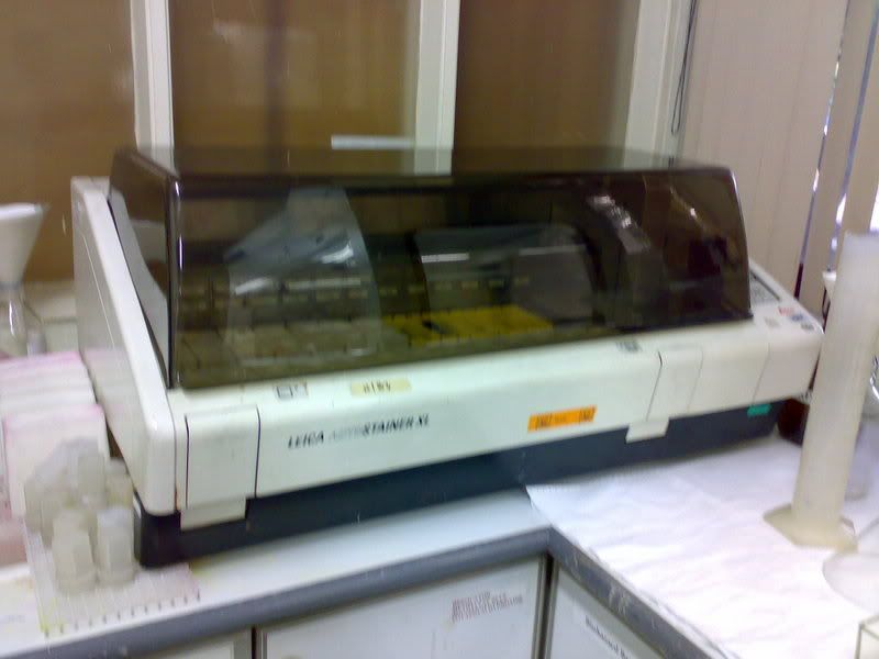
For routine H&E staining, the process is fully automated with the help of this machine (Leica Autostainer XL):


Extracted from:http://www.spencerscicorp.com/html/slidestain.html

This is how it works: After loading the rack into the machine, press LOAD and check that the machine is programmed to routine H&E staining. After that, the whole staining process will be carry out by the machine in this format:

1) Xylene--> 2 minutes

2) Xylene--> 2 minutes

* Steps 1 & 2- remove wax that has not melt off as wax are not water soluble.

3) Abs alcohol--> 1 minute

4) 95% alcohol--> 1 minute

5) 70% alcohol--> 1 minute

*Steps 3-5- remove xylene and rehydrate the specimens

6) Water--> 1 minute

*As haematoxylin (Hx) is aqueous based, must wash with water first

7) Hx--> 4 minutes

8) Hx--> 3 minutes

* Steps 7 & 8- stain the whole specimen 9)

9) Water--> 1/2 minute

10) 0.5% acid alcohol--> 2 dips

* Differentiation- remove excess Hx
11) Water--> 1 minute

12) Lithium carbonate--> 2 minutes

* An alkali which blue the specimen when place in water

13) Water--> 2 miuntes

* Blue in water

14) Water--> 3 minutes

15) Eosin--> 1/2 minutes

* Stains cytoplasm

16) 70% alcohol--> 1 minute

17) 95% alcohol--> 1 minute

18) 4 rounds of abs alcohol--> each for 1 minute

* Steps 16-18- dehydration as water cannot mix with xylene)

19) 3 rounds of xylene--> each for 1 minute

* Remove alcohol as depex used for mounting is xylene-based and also to raise refractive index of tissue

Results:

  • Nucleur component- blue
  • Cytoplasmic component- red

After the above process, the specimens will be send for mounting which is also automated.

Special staining

Principle: Some cases need special stains for diagnosis. H&E is not able to demonstrate the desired components.

Types of special stains:

  • Periodic Acid Schiff

  • GMS Fungus

  • Reticullum ll

  • Alcian Blue

  • Iron

  • Giemsa

  • Congo Red

  • Gram
Procedures:
1) Print barcode label(s) (according to biopsy number assgined to the specimen)
2) Paste lable(s) on the frosted end of the slide
3) Dewax and hydrate slide
4) Load the slide covered with special wash solution to prevent the specimen from drying up into the instument
5) Load appropriate stain kit into the reagent carousel (depending on the type of stains)
6) Select Run
* Pre-checklist will appear. Ensure all slides and reagent kit are in place. Cap of reagent kit must be open.
7) Enter number of slides and hit the Run button
The machine will now initiate the staining by reading the slide and reagent barcode labels. After the barcodes are read, information for the staining will be 'downloaded' from the NexES computer to the staining module and the run will resume. When the run has completed, click 'SIGN OFF' and remove the slides from the instument. Drain slides then load into rack and place in 95% alcohol to remove residual liquid before dehydration, clearing and mounting.

Manual Staining
Principle: The Ventana NexES machine are not able to carry out some special stains. An example is the Ziehl Neelsen staining. (I only perform this manual staining so far)

Tuberculosis Test/ Ziehl Neelsen:
Principle:To demomonstrate acid fast bacteria belonging to the genus mycobacterium responsible for causing tuberculosis. (diagnosis test for tuberculosis)

Control: Any tissue containing acid fast organism.

Reagents required:
  • Commercial TB colour carbo reagent--> stains tubercle bacilli red
  • Loeffler's Methylene Blue--> background staining
  • 1% potassium hydroxide
  • 1% acid alcohol

    Procedures:
    1) Dewax and bring sections to water
    2) Stain with commercial TB colour carbo reagent for 5 minutes
    3) Wash in running water
    4) Differentiate with 2 rounds of 1% acid alcohol until colourless (patient is negative), light pink (patient is positive)
    5) Wash in water
    6) Counterstain with 1% Loeffler's methylene blue for 10-15 seconds
    7) Wash in water and go to 95% alcohol to control intensity of the blue colour
    *if very blue, go to 70% alcohol (the more diluted the alcohol, the greater the differentiation)
    8) Check under microscope, if too much blue has gone off due to differentiation, repeat steps 6&7
    9) Dehydrate in absolute alcohol, clear in 3 rounds of xylene and mount

    Results:
  • Positve- red tubercle bacilli on blue background
  • Negative- sky blue

Finally, its the end of my long posting. LOL. Feel free to ask me any easy questions. Hope you guys continue to learn more from SIP! Take care.=)

June Tham
TG02
0505073G


Saturday, July 21, 2007

Cytology Pictures

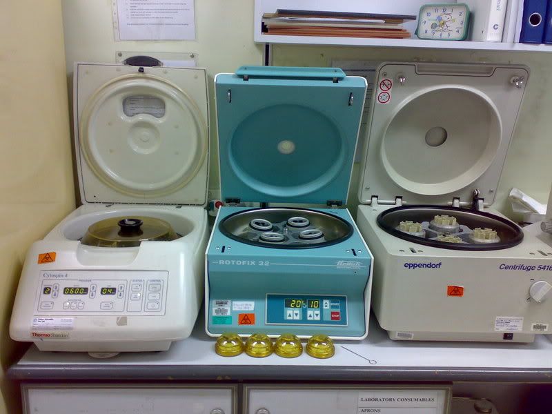
Centrifugers

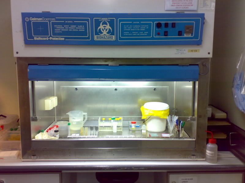
Safety Cabinet Level 2 b

Safety Cabinet Level 2 b

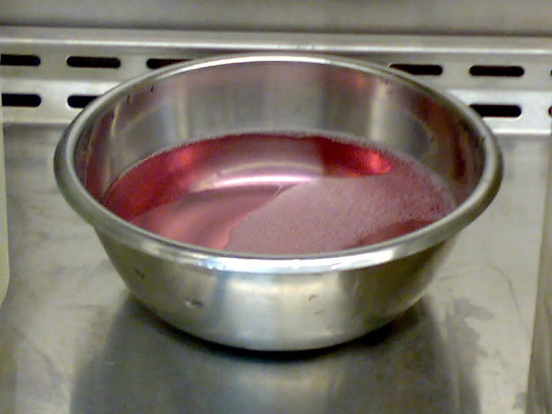
Virkon solution

Autostainer

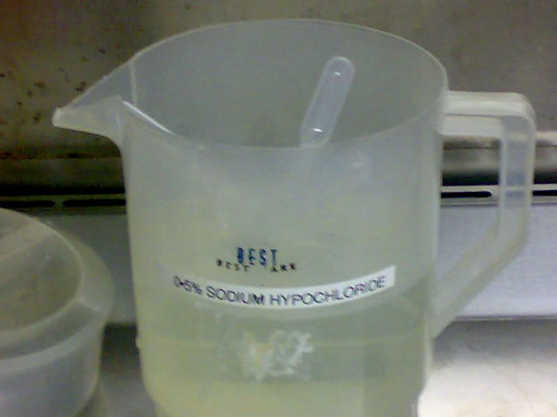
0.5% Sodium Hypochloride

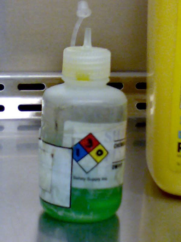
Shandon reagent

Oven

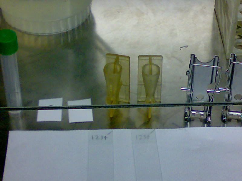
Cytocentrifuge funnel, clamp, filter paper and labelled glass slides

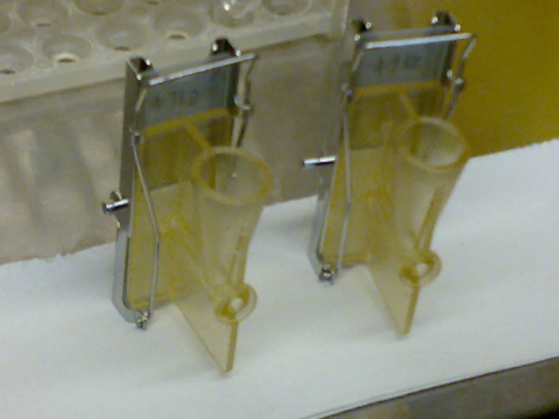
Cytocentrifuge funnel, clamp, filter paper and labelled glass slides ready for cytocentrifugation


Disclaimer: These pictures were taken by me with permission from my supervisor; the latter has seen each picture and allowed me to use them to enhance the learning process.

Student Internship Programme - Cytology

Hey people, Desmond here. I've been attached to the cytology for 4 weeks now and have been doing non-gynaecological procedures for the first 3. In this posting, I will guide you through what is done from the time a urine specimen is recieved right until it is "reported" to the cytologist.

I will be posting pictures soon and feel free to ask any EASY questions =].

Subject title
Pathology (Cytology)

Aims
Cytology testing of Urine for malignancy

Introduction
The principle of cytology testing of urine is to differentiate the nucleus and cytoplasm of the cells to detect malignancy by comparing the Nucleus: Cytoplasm ratio (N:C ratio). The Papanicolaou staining method is used for this. It is a polychrome staining reaction (staining the cytoplasm of different cells different colours) designed to exhibit differences in cellular morphology, maturity and metabolic activity. Because intact cells in a cytological smear tend to overlap and some appear in 3D configurations, the greatest value of the Pap staining method are the resultant transparency of he cells and clear definition of nuclear detail.

Urine however has small number of cells; Cytocentrifugation is needed to pull the cells into a confined/defined region on the glass slide.

Materials
Glass slides
Depex
Xylene
Autostainer
Centrifuge
Cytocentrifuge
Cytocentrifuge funnel and clamp
10ml centrifuge tube
22mm by 22mm coverlips
Labels/Stickers
Shandon reagent
0.5% sodium hypochloride
Virkon solution

Methods
(a) Receiving Specimens (Urine)

1. Scan the time onto the patient’s form
2. Fill in the type of specimen and initials of the person receiving the specimen (e.g. DH)
3. Label the patient’s form with pre-made stickers which are chronologically numbered and bar-coded.
4. Assign a cytologist to the case and record details like case number and the cytologist assigned to the case.

(b) Processing specimen (urine)

1. Enter Biosafety Cabinet Level 2.
2. Remove the container with the patient’s urine from the biohazard bag
3. Compare IC numbers on patient form and the container
4. Label the container and a 10ml centrifuge tube with the pre-made stickers (e.g. NG1234/07)
5. Indicate on the patient’s form the following:
Volume of urine
Condition of urine (e.g. Colourless, bloody or yellowish)
Initials of the person processing the urine
Date
Number and type of stains required (in this case 2 Pap stains)
6. Gently mix the urine by overturning the container
7. Aliquot about 10ml of urine into the 10ml centrifuge tube
8. Centrifuge the urine for 10 minutes at 2000rpm.
9. Prepare 2 labelled glass slides for cytocentrifugation
10. Decant the supernatant into 0.5% Sodium Hypochloride
11. Add 6 drops of Shandon reagent to the cell pellet.
12. Mix by pipetting up and down
13. Add 3 drops of the mixture of cells and Shandon into the cytocentrifuge funnel.
14. Cytocentrifuge for 6 minutes at 800rpm
15. After cytocentrifugation, fix the glass slides in 95% alcohol.

(c) Staining

1. After fixation for 20 to 30 minutes, load the glass slides onto a staining rack.
2. Load the rack into the autostainer
3. Set the autostainer to the Papanicolaou staining programme.

(d) Mounting

1. After staining, mount the 2 glass slides.
2. Dip the glass slide into xylene
3. Add a drop of depex to the stained side of the glass slide
4. Place a coverlip on the Depex
5. Press out any air bubbles
6. Place the glass slides into an oven (45 degrees Celsius) to let the Depex solidfy (5 to 10 minutes)

(e) Microscopic observation and reporting

1. Observe under microscope to determine if there is an adequate number of cells or staining for the cytologist to detect any abnormalities/malignancy

2. Report the case including the patient’s form and glass slides to the cytologist to detect abnormalities (malignancy)

Results
(Reference)
Nuclei – Blue
Acidophillic cells – Red
Basophilic cells – Blue-green
Erythrocytes – Orange-red

(Urine tested)
Nucleus appears very large when compared to cytoplasm. There are multinucleated cells and the nuclei appear to be intensively stained blue-green.

Discussion

The patient’s Cells are malignant. There is nuclear enlargement without an increase in the overall size of the cell, giving a decreased cytoplasm to nuclear ratio. There is also hyperchromasia due to increased amounts of DNA. The nuclear outline appears irregular and there is a variation in size and shape. Moreover, nucleoli increase in size and number. Abnormal cell division results in multinucleation. Finally, there is uneven distribution and variation in size and chromatin particles.


Desmond Heng Chih Pheng


0503179D


TG02

Thursday, July 19, 2007

Long-awaited answers!

....

Despite my despairing pleas, everyone didn't heed them and -still- posed tough questions for me! sigh.. and here goes,


Phui Yuen: hahaha yeah countless PBFs done, but there's still room for improvement! regarding your qns 'under what disease/clinical presentation is ESR done? or that ESR is just a general routine test done?', ESR isn't considered a general routine test but a miscellaneous(single) test ordered separately by the consultant himself/herself. ESR, as stated, is a nonspecific screening test whose results do not confirm a diagnosis of diseases. for eg, a patient can have a normal ESR readings and still have a problem. so yeah, basically it's up to the consultant to decide if he/she wants the test done to aid in diagnosis but that will be more towards clinical aspect.


Azhar: 'Since you said that the erythrocyte sedimentation process can be affected by a number of factors, is there any special equipment that is used to minimise these factors?'

hm actually there isn't any special equipment. perhaps the special and most important one is the ESR stand to support the tube in a vertical position for a more reliable and accurate reading. if you had noticed in the factors that i've stated, most of them would largely lean towards human variation(errors). also, the ESR stand is secured at the middle of the table and not near the ends where the alignment may not be equal and certainly not near any centrifuges.


Eugene & Nisha: hahaha. 'what's hairy cells?' they are cells with 'hairs'. ok lame :P the image is taken from a previous control slide but the presence of hairy cells in the blood basically shows that the patient is suffering from hairy cell leukemia(hcl), and hcl is classified under chronic lymphoid leukemia and a b-cell disease. the abnormal b-cell will have hairlike cytoplasmic projections on its surface. i dare not go into details for i'm not too sure myself!


Doreen: yeah my lab uses hematek2000 stainer but occasionally, manual staining using leishman stain. that's during time constraints. as for FBC report reflecting low platelet count, the follow-up actions will be to check for fibrin clot in the sample using wooden applicator sticks, any presence of giant platelets or platelet clumps in the blood film diagnosis which will be screened by the senior med techs and after which, they can proceed by doing manual differential count when necessary and report significant RBC and platelet findings.


Yeng Ting: to your first qns 'Rouleaux formation is the stacking of erythrocytes? Is the settling of erythrocytes equal to rouleaux formation?' yes, rouleaux formation is the stacking of RBCs atop one another. in ESR, the basic principle of the test is for the RBCs to form rouleaux and as the complex gets 'heavier', it generally settles down into what we call the packed cell volume. to your second qns 'In what ways can the ESR results be affected?' let me put this simply with a patient with sickle cell anemia as an example, sickle cells unlike normal RBCs are shaped like crescents. these sickle cells will still settle down but unable to form a typical rouleaux like normal RBCs probably with 'gaps' between them. so, going back to the principle of ESR, results will be affected by the structural variations of cells and in this case, give a decreased value.


Ying Ying: hahaha. i didn't see this coming. 'Why perpendicular and not vertical?' define perpendicular and it has the meaning of being vertical and upright in the sense that the 90-degree angle is created by the table surface and the ESR tube. so, simply put it this way, perpendicular in this case equals vertical!


Eunice: !!! 'Why are there 2 reference ranges for females?' the reference ranges listed were obtained locally based on results of healthy individuals. there being 2 ranges for females divided at about 50 years are probably due to normal physiology of females like menopause etc. there are significant changes of the mean result of females at <50>51 years during the tabulation of data and so the reference ranges were divided as so for females.


Lizzie: really? your place received over a thousand samples for ESR?! wow. hahaha.

ESR here are done manually. whether it is done before or after FBC totally depends on the amount of blood sample received. ideally, it would be done before FBC due to the waiting time of an hour but if there aren't enough blood in the sample to run both FBC and ESR, running FBC will be the priority.


Chaur Lee: yeah there certainly will be interference of results after 60 minutes as RBCs will still continue to settle down beyond the time frame resulting in false high readings. here, the count-down timer is a med tech's best friend. the reading is taken plus/minus 1 minute of the 60 minutes when the alarm sounds, so there is virtually a remote possibility that these results are not used. however, it's human to err so you wouldn't want to discount human errors if it happens? and yup, variations in RBC affect ESR results but in this case, ESR being a miscellaneous test is ordered by the consultant so maybe it's fair to say that we do our jobs by performing the test and report the results. :P


Wing Fat: 'What is platelet satellitism?' i must admit this is a new term to me when i saw the control slide and took a pic to share with you guys! how could you do this to me.. but yeah after some research(thanks ah), platelet satellitism is characterized by platelets forming around polymorphonuclear leucocytes(neutrophils) and are seen in blood films stained by Wright's stain prepared from EDTA-anticoagulated blood samples and not seen in other anticoagulants like sodium citrate, heparin etc. wouldn't go into details as this is only what i've learnt! thanks ah wing fat.


Jiaxin: hahaha. i suppose if the company is paying for your living, it is a must to learn to do everything. us as interns, if the supervisor deems you fit as to run patient samples, you are being treated like one of them and will be good to help around to your abilities and learn at the same time. so, the answer to your question is DO EVERYTHING!


Juexiu: for panic alarms such as excessively abnormal levels of platelets, haemoglobin and total white, we have to inform the wards immediately. while for general abnormal results from tests, we report the results and include comments with it.


Ci Liang: yeah, variations in RBC sizes affect the ESR results. i asked a senior med tech and she told me ESR for children were done mostly done in the western countries and it had been done here in the past, but not at present. but i've also found a reference range for children of 0-10mm/hr developed at Baptist Memorial Health Care Corporation, Memphis, Tennessee. hopefully it helps. :D


Peishan: this question got me thinking for quite a while, and i actually set up an experiment today just for you! it is definitely important to mix the ESR tube thoroughly before setting the test up to achieve a state of homogeneity between the components of blood. if blood is not mixed well, RBCs will settle at the bottom and form 2 clear distinct layers of plasma and RBCs. from what i've observed from my experiment, setting up the ESR test on blood that was not mixed(tube 1) will therefore allow the plasma region to be catapulted right to the top of the tube in place of homogenized blood from the control(normal ESR/tube 2). from the start, in tube 1, it can be observed that it isn't a fair test due to the segregation of plasma and RBC layer giving a reading of about 8mm following by the RBC layer that is unevenly spread while in tube 2, the reading is at the zero mark. after 60 minutes, the readings which i recorded were 85mm/hr and 32mm/hr in tubes 1 and 2 respectively. this clearly shows that the tube without mixing gives a false high result. after discussing on the findings with a senior med tech, it can be said that in the tube 1, some areas of the red cell layer had already settled down which MAY had formed rouleaux. with this uneven distribution of weight in the vertical ESR tube, say at the centre, it may force the RBCs further down along with gravitational pull which will give a high reading come the end of 60 minutes. these are of course, based on my observations and discussion, i really hope you did understand what i'm trying to bring. perhaps mr alvin poh[if he does read] can explain why?


Joan: both primary and secondary controls are commercial controls, given by the manufacturer of the automation. in the morning, the 3 levels of control (low/medium/high) is run in the machine before running patient samples to ensure that results fall within the manufacturer's given reference ranges. running secondary control also utilises the commercial controls but the difference is that only 1 of the 3 levels are used at the given time intervals. this is part of internal quality control and it also functions to keep patients' results in check within the reference ranges and that results are quality-assured.


Sharifah: hahaha in the lab, the ESR station is secluded in the middle of a single table. only materials involved in ESR are placed there, namely ESR stand, ESR tubes, timer, record book etc. the station is usually left alone after setting the ESR tests and no other materials known to cause vibrations are placed there. the only vibrations then, can only be due to people treating the table like a drum! but trust me, why play at the station? it's no fun! :P


AHHHHHHHHH! and trust me, answering questions are no fun! but of course, end of the day, i hope i did help in easing those lingering doubts that you once had and learnt something from my experience!

case closed! *wide grins :D* till my next post.. have fun everyone!

Saturday, July 14, 2007

Peek-tures

Mixing of blood to prevent clotting


Patient with chronic granulocytic leukaemia
(Note the huge amounts of total white cells
as well as different stages of differentiating cells)


Hairy cells


Platelet satellitism
(Clumps of platelets adhering to neutrophils)


ESR station


Images released with the permission of company supervisor

- Alex