Monday, December 10, 2007

Particulars of patient

Name:Khong Fay Seah
Sex:Female
Age:27
Clinical Diagnosis
Complaints: fever,chills,dysuria
Diagnosis:urinary tract infection
Antibiotic treatment: if any (nil)


Possible Microorganisms

Description

Investigations

Escherichia coli(Most common cause of UTI)


-Belongs to family of Enterobacteriaceae(Escherichia)

-Clinical findings: UTI (Pyelonephritis and Cystitis), diarrhea disease

-Is a member of normal intestinal flora

-Microscopy and gram staining: Gram negative rod

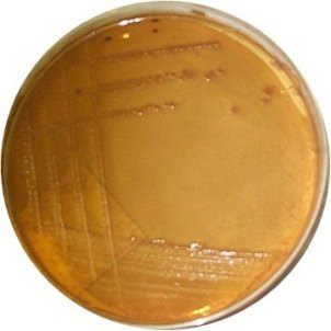
-Culture: Haemolyic colonies on blood agar, lactose fermenting colonies (Red/Pink) on MacConkey agar, green metallic sheen on EMB agar

-Biochemical tests:
-Indole positive
-Methyl red positive
- Voges-Proskauer (VP) negative
-Urease negative
-Citrate negative
-Catalase positive
-Oxidase negative
-TSI: acidic slant/acidic deep/no H2S

-Susceptibility testing: ampicillin, cephalosporin, aminoglycosides, sulfonamides

Staphylococcus saprophyticus(Second most common cause of UTI)


-Belongs to family of staphylococci

-Clinical findings: Urinary tract infection

-Present in the urinary tract and bladder of sexually active females

-Microscopy and gram staining: Gram positive cocci in clusters

-Culture: Pale colonies on blood agar

-Biochemical tests:
-Catalase positive
-Coagulase negative

-Susceptibility testing:Quinilone

Klebsielle Pneumoniae


- Belongs to family of Enterobacteriaceae(Klesbsiella)

- Clinical findings: urinary tract infection

-Present in the respiratory tract and feces of about 5% normal individuals

-Microscopy and gram staining: Gram negative rod

-Culture: Pink, mucoid colonies on MacConkey agar

-Biochemical tests:
-Indole negative
- Methyl red negative
-Voges-Proskauer reaction positive
- Urease negative
-Citrate positive
- TSI: acidic slant/acidic deep/no H2S

-Susceptibility testing:Cephalosporin

Enterobacter aerogenes


- Belongs to family of Enterobacteriaceae(Enterobacter)

- Clinical findings: urinary tract infections and sepsis

- Present in the intestinal tract-

-Microscopy and gram staining: Gram negative rod

-Culture:Pink to purple colonies on EMB agar

-Biochemical tests:
-Indole negative
-methyl red negative
-VP positive
-urease negative
- Citrate positive
-TSI: acidic slant/acidic deep/no H2S

Susceptibility testing: cephalosporin

Proteus mirabilis


- Belongs to family of Enterobacteriaceae(Proteus)

- Clinical findings: urinary tract infections and produce bacteremia, pneumonia

-Microscopy and gram staining: Gram negative rod
-Culture: Pale colonies on MacConkey agar, swarming growth on blood agar

-Biochemical testing:
-Indole negative
-Methyl red positive
-VP positive/negative
-Urease positive
- Citrate positive
-Oxidase negative
-TSI: alkaline slant/acidic deep/H2S

-Susceptibility testing: penicillin, cephalosporins, quinolones

Enterococcus faecalis


- Belongs to family of Streptococci

-Clinical findings: abdominal abscess, urinary tract infection ((bladder infection, Pyelonephritis)

-Present in colon

-Microscopy and gram staining: Gram positive cocci in chains

-Culture: Red dot colonies on MacConkey agar

-Biochemical testing:
-Decolourise the litmus milk,
-Indole negative
-Catalase negative

-Susceptibility testing: Vancomycin,ampicillin

Pseudonomas aeruginosa


- Belongs to family of Pseudomonads

-Clinical findings: Typically infects the pulmonary tract, urinary tract(Pyelonephritis)

-Distributed in nature and is commonly present in moist environments in hospital

-Microscopy and gram staining: Gram positive spherical

-Culture: Greenish pyocyanin pigment on MacConkey agar

-Biochemical testing:
-Indole negative
-Urease negative
-Oxidase positive,
-TSI: allaline slant/alkaline deep

-Susceptibility testing: Ciprofloxacin,imipenam


PICTURES OF THE POSSIBLE CAUSATIVE AGENTS


Photo Sharing and Video Hosting at Photobucket


E Coli ( red dot colonies on Mac agar)

Photo Sharing and Video Hosting at Photobucket

K. Pneumonia (red/pink colonies on Mac)

Photo Sharing and Video Hosting at Photobucket

Staphyloccocus Saprophyticus
Photo Sharing and Video Hosting at Photobucket
Pseudomonas aeruginosa
Photo Sharing and Video Hosting at Photobucket
Enterococcus faecalis
Photo Sharing and Video Hosting at Photobucket
Enterobacter aerogenes
Photo Sharing and Video Hosting at Photobucket
Proteus mirabillis
References

http:www.yahoo.com>image search> Proteus mirabilis
http:www.yahoo.com>image search> Enterobacter Aerogenes
http:www.yahoo.com>image search>Enterococcus faecalis
http:www.yahoo.com>image search>Pseudomonas aeruginosa
http:www.yahoo.com>image search>Staphyloccocus Saprophyticus
http:www.yahoo.com>image search>K. Pneumonia
http:www.yahoo.com>image search> E Coli
http://www.mc.maricopa.edu/~johnson/labtools/Dbiochem/emb.html>EMB agar
http://en.wikipedia.org/wiki/MacConkey_agar> MacConkey Agar
http://en.wikipedia.org/wiki/Blood_agar#Blood_agar_types> blood agar
Book
Tony Hart, Paul Shears (2004).Color Atlas of Medical Microbiology: Elsevier’s Health Sciences Department


kai lin
0503211E

Sunday, December 9, 2007

MMIC dPBL Case 3 (Maisy Hong - 2nd posting cont)

Pictures of possible causative agents of case 3

Pseudomonas aeruginosa growing on XLD agar Acinetobacter baumanii as seen under microscopy
Serratia marcescens growing on XLD agar
Candida albicans as seen under microscopy

Staphylococcus saprophyticus growing on nutrient agar Enterococcus faecalis as seen under scanning electron microscopy
Escherichia coli as seen under microscopy
Proteus mirabilis growing on XLD agar

Klebsiella pneumoniae as seen under microscopy
References:
All images are credited to
1) www.yahoo.com > image search > "microorganism name"
2) www.wikipedia.org > "microorganism name" > search
Information are credited to
3) Murray, P.R., Kobayashi, G.S., Pfaller, M.A., Rosenthal, K.S. (1994). Medical Microbiology Second Edition. London: Mosby-Year Book, Inc
4) Medical Microbiology Notes
5) www.yahoo.com > "microorganism name"
6) www.wikipedia.org > "microorganism name"
Tan Yi Wei Alex Tg02 0503222B

MMIC dPBL Case 3 (Maisy Hong - 2nd posting)

Learning issues:

1) Identify suspected microbial agents
2) Conduct self-directed learning based on specific case and symptoms
3) Propose relevant tests according to suspected causative agents
4) Propose treatment to suspected causative agents

Keywords: Female, 67 years, Fever, Chills, Bladder distension, Indwelling catheter, Urinary tract infection, Urine specimen

Patient's Particulars

Name: Maisy Hong
Age: 67 years old
Sex: Female

Clinical Diagnosis

Complaints: Fever, chills, bladder distension (inability to urinate)
Diagnosis: Urinary tract infection (UTI)
Note: On indwelling catheter (a urinary catheter is a plastic tube which is inserted through a patient's urinary tract into their bladder)

Diagnostic approach to UTI in adults

1) Patient symptomatic? Yes, go to 2a. No, go to 2b.
2a) Complicating factors? Yes, go to 3a. No, go to 3b.
2b) Asymptomatic bacteriuria
3a) Complicated UTI
3b) Recurrent episode? Yes, go to 4a. No, go to 4b.
4a) Recurrent UTI
4b) Symptoms of upper tract involvement? Yes, go to 5a. No, go to 5b.
5a) Pyelonephritis
5b) Consider cystitis, urethritis or vaginitis (lower tract)


Usually, before proceeding to microbial identification tests, a urinalysis and chemical dipstick testing will be performed on the urine sample and important factors of UTI such as leucocytes, erythrocytes, pH as well as presence of certain chemicals like nitrite are things to look out for. As we've covered this in clinical chemistry, I will just talk about the medical microbiological portion. :)


Below is a table of possible causative agents, characteristics, proposed tests for agent identification and antibiotic susceptibility testing (treatment).


(Please click the pictures for enlarged images)


Tan Yi Wei Alex Tg02 0503222B

CASE STUDY 4

Particulars of Patient
Name: Tong Wei Hong
Sex: Male
Age: 68 years old

Clinical Diagnosis
Signs and symptoms: Fever, chills, excessive phlegm, breathing problems Diagnosis: Bronchitis
Specimen collected: Sputum

Description of Bronchitis
Bronchitis is an inflammation of the bronchi (medium-size airways) in the lungs. Acute bronchitis is usually caused by viruses or bacteria and may last several days or weeks. Acute bronchitis is characterized by cough and sputum (phlegm) production and symptoms related to the obstruction of the airways by the inflamed airways and the phlegm, such as shortness of breath and wheezing. Diagnosis is by clinical examination and sometimes microbiological examination of the phlegm.

Chronic bronchitis is not necessarily caused by infection and is generally part of a syndrome called chronic obstructive pulmonary disease (COPD); it is defined clinically as a persistent cough that produces sputum (phlegm) and mucus, for at least three months in two consecutive years.

Possible Causative Agents

1. Adenovirus
Non-enveloped double-stranded linear DNA
Icosahedral nucleocapsid with a fiber protruding from each of the 12 vertices
Causes Bronchitis in the lower respiratory tract

2. Bordetella
Small, coccobacillary, encapsulated gram negative rod
Restricted to the respiratory tract (negative blood culture)

3. Haemophilus influenzae
Non-motile gram-negative coccobacillus
Found in the upper respiratory system of humans
Major cause of lower respiratory tract infections, associated with pneumonia

4. Moraxella catarrhalis
Gram-negative, aerobic, oxidase-positive diplococcus
May colonise and cause respiratory tract-associated infection in humans
Known to cause Bronchitis

5. Streptococcus pneumoniae
Gram positive lancet-shaped cocci
Arranged in pairs or short chains
Higher mortality in persons aged 65 and above


Investigations
The colour of the sputum may indicate the kind of microorganism in the sputum; yellow to green may indicate bacteria while white may indicate viruses. The sputum should be cultured to isolate the microorganisms listed above. In addition,

Biochemistry
-Gram staining
-Citrate test
-Catalase test
-Oxidase test
-Urease test

Microbiology
-Culture aerobically on blood agar plates

Antibiotic Suspectibility testing

Serology (Virus detection)
-Enzyme-Linked ImmunoSorbent Assay (ELISA)
-Haemagglutination


References
www.wikipedia.org > English > search > Bronchitis
www.wikipedia.org > English > search > Adenovirus
www.wikipedia.org > English > search > Bordetella
www.wikipedia.org > English > search > Haemophilus influenzae
www.wikipedia.org > English > search > Moraxella catarrhalis
www.wikipedia.org > English > search > Streptococcus pneumoniae


Desmond Heng
0503179D

Follow-up of case 6


Above is the procedure that i will carry out during laboratory to determine the identity of the microoraganism present in the vaginal swap.

Materials Needed:
1) Blood Agar
2) MacConkey Agar
3) Gram stain Reagent
4) Caoagulase Reagent
5) Oxidase Reagent
6) Catalase Reagent
7) Commercial ID Testing Kits
8) Biochemical Tests Reagents
9) Antibody lines
Patient: Ong Fei Fei
Age: 37 Years old
Complaints: Fever, pain during urination, virginal discharge
Diagnosis: UTI

1)Enterococcus Faecalis
Labortatory Diagnosis:
(i) Alpha, beta or non hemolytic colonies on blood agar
(ii) Grow in 6.5% NaCl and hydrolyzes esculin on the presence of 40% bile
(iii)Catalase Negative

2)Eschericahia coli
Labortatory Diagnosis:
(i) Lactose fermenting on macConkey agar
(ii) TSI show acid slant, acid butt with gas but no H2S
(iii)Indole +
(Iv) Motile

3)Enterobacter-Klebsiella-Serrtia Family
Labortatory Diagnosis:
(i) Lactose fermenting on macConkey agar
(ii) Appear as mucoid colonies on agar plate

4)Proteus-Providencia-Morganella Family
Labortatory Diagnosis:
(i) Non-lactose fermentor on Macconkey
(ii) Distinct swarming on blood agar
(iii)P.vulgaris and P.mirabilis produce H2S that blacken the butt of TSI agar
(iv) P.mirabilis is indole negative whereas the other 2 are indole positive
(v) Urease Positive

Antibiotic suceptibility Testing will be carried out on antibodies mention in previous posting after bacterial is identified.

Yeo Ching Wei
0503288C

MMIC Blog Post 2

Patient’s Particulars:
Name: Kwan Siew Yan
Sex: Female
Age: 29 years

Clinical Diagnosis:
Complaints: Diarrhea
Diagnosis: Entercolitis
Antibiotic Treatment: Nil

Possible organisms:
1) Vibrio species:
V. parahaemolyticus- Facultative anaerobes
Microscopic features:
· “Curved”, comma-shaped rods
· Motile with polar flagella
Laboratory investigations:
· Gram staining: Negative
· Oxidase: Positive on blood agar
· TSI: acid slant/acid deep without H2S production
· 0/129 (Vibriostat) Disk: Susceptible
· Nutrient Broth with 0% or 6-8% NaCl: Requires 1-7% NaCl
· Decarboxylase: Positive
· Culture media: TCBS as green colonies due to absence of sucrose fermentation
Antibiotic-susceptibility test:
· Tetracycline

2) Campylobacter species:
C. jejuni- Microaerophilic
Microscopic features:
· “S” shaped rods
· Darting motility
Laboratory investigations:
· Gram staining: Negative
· Oxidase: Positive
· Catalase: Positive
· TSI: Alkaline slant/alkaline deep
· Hippurate hydrolysis: Positive
· Culture media: Campylobacter agar incubated under microaerophilic conditions at 42oC as colourless or gray colonies
Antibiotic-susceptibility test:
· Erythromycin

3) Clostridium species:
C. perfringens- Obligate anaerobes
Microscopic features:
· Large, motile rods
Laboratory investigations:
· Gram staining: Positive
· Hemolysis test: Beta-hemolysis on blood agar as flat, spreading, translucent colonies
· Nagler test: Lecithinase positive
· Thioglycolate medium: Various sugars present
· Action on milk: Turned acid or digested
· Culture media: Egg yolk agar as colonies surrounded by precipitates with absence of spores
Antibiotic-susceptibility test:
· Penicillin

4) Escherichia species (Diarrheal diseases) – Facultative anaerobes
Microscopic features:
· Rod-shaped
Laboratory investigations:
· Gram staining: Negative
· TSI: Acidic slant/acidic deep with gas
· Indole: Positive
· Methyl red: Positive
· Voges-Proskauer: Negative
· Citrate: Negative
· Urease: Negative
· Culture media: EMB agar as greenish metallic sheen or MAC agar as red or pink colonies
Antibiotic-susceptibility test:
· Ampicillin

5) Salmonella species- facultative anaerobes:
Microscopic features:
· Rod-shaped
· Motile
Laboratory investigations:
· Gram staining: Negative
· TSI: Alkaline slant/acidic deep with H2S production
· Indole: Negative
· Methyl-red: Positive
· Voges-Proskauer: Negative
· Citrate: Positive
· Urease: Negative
· Culture media: EMB agar as translucent colonies or MAC agar as uncoloured colonies (non-lactose fermenting colonies)
Antibiotic-susceptibility test:
· Ampicillin

6) Shigella species- facultative anaerobes:
Microscopic features:
· Slender, rod-shaped
· Non-motile
Laboratory investigations:
· Gram staining- Negative
· TSI: Alkaline slant/acidic deep without gas or H2S production
· Indole: Negative
· Methyl-red: Positive
· Voges-Proskauer: Negative
· Citrate: Negative
· Urease: Negative
· Culture media: Culture media: EMB agar as translucent colonies or MAC agar as uncoloured colonies (non-lactose fermenting colonies)
Antibiotic-susceptibility test:
· Ampicillin

Pictures of possible organisms:
1) V. parahaemolyticus on TCBS agar


2) C. jejuni


3)Enteroinvasive E. coli (EIEC)
  • EMB agar

  • MAC agar


4) Salmonella species
  • EMB agar


5) Shigella species
  • MAC agar


References
1) http:www.yahoo.com>image search> Vibrio parahaemolyticus
2) http:www.yahoo.com>image search> Campylobacter jejuni
3) http:www.yahoo.com>image search> Enteroinvasive E. coli
4) http:www.yahoo.com>image search> Salmonella
5) http:www.yahoo.com>image search> Shigella


Tham Wan Jin June
TG02
0505073G

Flowchart of Identity Testing - Group post 1


7 Biochemical Identity Tests

(1) Kligler Iron Agar Test
It is used primarily to determine the carbohydrate fermentation and hydrogen sulphate production.
Slant
Butt
Acid Yellow
Acid Yellow
Alkaline Red
Alkaline Red

Red slant/ yellow Butt = Glucose fermented
Yellow slant/ yellow butt = Glucose fermented + lactose fermented
Red slant/ red butt = neither of them fermented

(2) Simon Citrate Agar Test
It is used to determine if an organism is capable of using citrate as sole source carbon for metabolism and growth
Positive
Growth with/ without an intense blue colour on the slant
Negative
No growth, no colour change

(3) Motility Test
It is used to determine if a microorganism is motile.

(4) Urea Hydrolysis Test
It to detect urease activity of both rapidly urea positive organism as well as a number of enterobacteriacae
Positive
Pink colour on the slant
Negative
No change in colour (pale yellow)

(5) Phenylalanine Deaminase Test
It is to determine the ability of organism to deaminate phenylalanine to phenylpyruvic acid
Positive
Light to dark green on the slant
Negative
No change in colour (yellow)

(6) Malonate Test
It is to determine the ability of organism to use malonate as a source sole of carbon
Positive
Light Blue to deep blue throughout the medium
Negative
No change in colour

(7) Indole Test
It is to determine the ability of organism to hydrolyze and deaminate tryptophan with the production of indole, pyruvic acid and ammonia
Positive
Red halo ring seen
Negative
No change in colour (yellow)

MMIC 2ND POSTING: CASE 5

CASE STUDY 5

Particulars
Patient: Wong Fei Hong
Sex: Male
Age: 37
Clinical Diagnosis
Complaints: Fever, swelling around operation wound
Diagnosis: wound infection
Antibiotic treatment (if any): Nil

Specimen Particulars
Specimen: Specimen 5 (Swab)
Date of collection: 26/11/2007
Time of Collection: 9 am

Result of First and Second Investigation through Gram Staining and Culturing


Biochemical Tests and Antibiotics Susceptibility Testing


Pictures of the Listed Microorganisms
  • Staphylococcus aureus


Left: Staphylococcus aureus gram stain
Right: Staphylococcus aureus on blood agar

  • Streptococcus pyogenes


Left: Streptococcus pyogenes gram stain
Right: Streptococcus pyogenes on blood agar


  • Enterococci


Left: Enterococci gram stain
Right: Enterococci on blood agar


  • Clostridium perfringens


Left: Clostridium perfringens gram stain
Right: Clostridium perfringens on blood agar

  • Pseudomonas aeruginosa


Left: Pseudomonas aeruginosa gram stain
Right: The soluble blue pigment pyocyanin is produced by many strains of Pseudomonas aeruginosa


References


Book:
Medical Microbiology, 23rd Edition Warren Levinson

Website:
http://keprice.myweb.uga.edu/pseudomonas_files/image002.jpg

http://www.bmb.leeds.ac.uk/mbiology/ug/ugteach/dental/tutorials/images/blood/staphaureus.jpg

http://www.textbookofbacteriology.net/pseudomonas.html

http://www.textbookofbacteriology.net/normalflora.html

http://www.mgm.ufl.edu/~gulig/mmid/mmid-lab/labimage/spy1.jpg

http://biomarker.cdc.go.kr:8080/pathogenimg/plate/Streptococcus_pyogenes_blood_agar(colony-1).JPG

http://www.textbookofbacteriology.net/clostridia.html
http://www.genomenewsnetwork.org/gnn_images/news_content/01_02/Clostridium_seq/clost2.jpg


By:

Lin Juexiu

0503151C

Monday, December 3, 2007

Particulars of Patient
Name: Tong Wei Hong
Sex: Male
Age: 68 years old

Clinical Diagnosis
Signs and symptoms: Fever, chills, excessive phlegm, breathing problems Diagnosis: Bronchitis
Specimen collected: Sputum

Description of Bronchitis
Bronchitis is an inflammation of the bronchi (medium-size airways) in the lungs. Acute bronchitis is usually caused by viruses or bacteria and may last several days or weeks. Acute bronchitis is characterized by cough and sputum (phlegm) production and symptoms related to the obstruction of the airways by the inflamed airways and the phlegm, such as shortness of breath and wheezing. Diagnosis is by clinical examination and sometimes microbiological examination of the phlegm.

Chronic bronchitis is not necessarily caused by infection and is generally part of a syndrome called chronic obstructive pulmonary disease (COPD); it is defined clinically as a persistent cough that produces sputum (phlegm) and mucus, for at least three months in two consecutive years.

Possible Causative Agents
1. Adenovirus
-Non-enveloped double-stranded linear DNA
-Icosahedral nucleocapsid with a fiber protruding from each of the 12 vertices
-Causes Bronchitis in the lower respiratory tract

2. Bordetella
-Small, coccobacillary, encapsulated gram negative rod
-Restricted to the respiratory tract (negative blood culture)

3. Chlamydia pneumoniae
-Obligate intracellular bacteria
-Require host cells for growth

4. Moraxella catarrhalis
-Gram-negative, aerobic, oxidase-positive diplococcus
-May colonise and cause respiratory tract-associated infection in humans
-Known to cause Bronchitis

5. Streptococcus pneumoniae
-Gram positive lancet-shaped cocci
-Arranged in pairs or short chains
-Higher mortality in persons aged 65 and above

Desmond Heng

0503179D

TG02


MMIC POST: CASE 5

MEDICAL MICROBIOLGY: CASE STUDY 5

Particulars
Patient: Wong Fei Hong
Sex: Male
Age: 37

Clinical Diagnosis
Complaints: Fever, swelling around operation wound
Diagnosis: wound infection
Antibiotic treatment (if any): Nil

Specimen Particulars
Specimen: Specimen 5 (Swab)
Date of collection: 26/11/2007
Time of Collection: 9 am

Step 1
Microscopy Examination
ž Gram stain is to be order.
ž This is to identify the gram reaction (purple-blue indicate gram-positive; pink indicate gram-negative) and morphology (shape: cocci, rods, bacilli and others) of the bacteria.

Step 2
Culture
ž Two types of culturing condition: aerobic and anaerobic
ž Select appropriate culture media
ž For wound infection as there is common media used.

Step 3
Biochemical tests which is to identify the microorganisms family
ž Tests to use will depend on the morphology of the bacteria.

Step 4
Antibiotic Susceptibility
ž Choose the appropriate antibiotic for choosing the correct antibiotic for the patient.


Possible Microorganisms


Antiobiotics Used


Lin Juexiu

0503151C